Download
1 / 37
410 likes | 744 Views
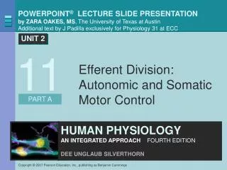
Efferent Division: Autonomic and Somatic Motor Control. 11. About this Chapter. Autonomic division Antagonistic controls Somatic motor division CNS control of skeletal muscles through neuromuscular junctions ****** Review the overall organization of the NS. Autonomic Division: Homeostasis.

E N D
About this Chapter • Autonomic division • Antagonistic controls • Somatic motor division • CNS control of skeletal muscles through neuromuscular junctions ****** Review the overall organization of the NS
Autonomic Division: Homeostasis • Antagonistic branches • Parasympathetic • “Rest and digest” • Restore body function • Sympathetic • “Fight or flight” • Energetic action
Autonomic Division: Homeostasis Figure 11-1
Autonomic Pathways • Coordination of homeostatic responses • Autonomic • Endocrine • Behavioral Figure 11-2
Autonomic Control Centers • Hypothalamus • Water balance, temperature, and hunger • Pons • Respiration, cardiac, and urinary • Medulla • Respiration Figure 11-3
Two Efferent Neurons in Series Autonomic pathways Figure 11-4
Two Efferent Neurons in Series • CNS- tracts coming from brain to spinal cord • Preganglionic neuron- exits spinal cord and goes to ganglion • Ganglion- sympathetic chain ganglion runs along vertebral colum • Postganglionic neuron- runs down spinal nerve • Target tissue- can be muscle or gland
Sympathetic versus Parasympathetic Figure 11-7
Antagonistic Control Autonomic sympathetic and parasympathetic pathways Notice the mention of antagonistic responses & receptors involved Figure 11-5
III. Nerves- these are bundles of axons (nerve fibers) that are myelinated or unmyelinated. Each axon is surrounded by an endoneurium, groups of nerves are bundles into nerve fascicles surrounded by perineurium and the whole nerve is surrounded by epineurium. *Know the difference between neuron, nerve fiber, and nerve. Nerves are found in the PNS
Sympathetic versus Parasympathetic • Spinal cord exit • Neurotransmitters • Receptors
Autonomic Targets • Smooth muscle • Cardiac muscle • Exocrine glands • Endocrine glands • Lymphoid tissue • Adipose tissue
Autonomic Neuron Structure • Neuroeffector junction - synapse between a postganglionic autonomic neuron and target cell • Postganglionic axon - exits spinal cord to target cell • Varicosities - instead of axon terminals, there are multiple branches and varicosities along the axon over the surface of the target cell
Synapses in Autonomic Neurons • Neurotransmitter released to ECF • No synaptic cleft • Impact • Large area • Slow acting • Long duration
Review of Efferent Pathways AUTONOMIC PATHWAYS Adrenal sympathetic pathway Somatic motor pathway Parasympathetic pathway Sympathetic pathways CNS CNS CNS CNS ACh Adrenal cortex Nicotinic receptor KEY Ach = acetylcholine E = epinephrine NE = norepinephrine Adrenal medulla Ganglia E ACh Nicotinic receptor Ganglion NE ACh a receptor Muscarinic receptor Autonomic effectors: Blood vessel • Smooth and cardiac muscles • Some endocrine and exocrine glands • Some adipose tissue b1 receptor E ACh Nicotinic receptor b2 receptor Skeletal muscle Summary of the efferent pathways of the peripheral nervous system Figure 11-11
Review of Efferent Pathways Somatic motor pathway CNS KEY Ach = acetylcholine E = epinephrine NE = norepinephrine ACh Nicotinic receptor Skeletal muscle Figure 11-11 (1 of 5)
Review of Efferent Pathways AUTONOMIC PATHWAYS Somatic motor pathway Parasympathetic pathway CNS CNS KEY Ach = acetylcholine E = epinephrine NE = norepinephrine ACh Nicotinic receptor Ganglion ACh Muscarinic receptor Autonomic effectors: • Smooth and cardiac muscles • Some endocrine and exocrine glands • Some adipose tissue ACh Nicotinic receptor Skeletal muscle Figure 11-11 (2 of 5)
Review of Efferent Pathways AUTONOMIC PATHWAYS Somatic motor pathway Parasympathetic pathway Sympathetic pathways CNS CNS CNS ACh Nicotinic receptor KEY Ach = acetylcholine E = epinephrine NE = norepinephrine Ganglia ACh Nicotinic receptor Ganglion NE ACh a receptor Muscarinic receptor Autonomic effectors: • Smooth and cardiac muscles • Some endocrine and exocrine glands • Some adipose tissue ACh Nicotinic receptor Skeletal muscle Figure 11-11 (3 of 5)
Review of Efferent Pathways AUTONOMIC PATHWAYS Somatic motor pathway Parasympathetic pathway Sympathetic pathways CNS CNS CNS ACh Nicotinic receptor KEY Ach = acetylcholine E = epinephrine NE = norepinephrine Ganglia ACh Nicotinic receptor Ganglion NE ACh a receptor Muscarinic receptor Autonomic effectors: • Smooth and cardiac muscles • Some endocrine and exocrine glands • Some adipose tissue b1 receptor ACh Nicotinic receptor Skeletal muscle Figure 11-11 (4 of 5)
Review of Efferent Pathways AUTONOMIC PATHWAYS Adrenal sympathetic pathway Somatic motor pathway Parasympathetic pathway Sympathetic pathways CNS CNS CNS CNS ACh Adrenal cortex Nicotinic receptor KEY Ach = acetylcholine E = epinephrine NE = norepinephrine Adrenal medulla Ganglia E ACh Nicotinic receptor Ganglion NE ACh a receptor Muscarinic receptor Autonomic effectors: Blood vessel • Smooth and cardiac muscles • Some endocrine and exocrine glands • Some adipose tissue b1 receptor E ACh Nicotinic receptor b2 receptor Skeletal muscle Figure 11-11 (5 of 5)
Visceral sensory neurons- Receptors in the viscera are free dendritic ends that send afferent signals caused by stretching, temperature and chemical changes, and irritation. Integration translates these signals into hunger, fullness, pain, or nausea. Visceral sensation may be hard to localize. Sometimes pain is called referred pain. A problem with an organ like the heart may send pain down the arm (not an area where the heart is located.) A map of referred pain: these are skin or body regions that present pain when there is visceral pain. The organ and site of referred pain are innervated by the same nerve. Referred Pain- Visceral Sensory Division
Somatic Motor Division • Skeletal muscle- target effector • Body movement- main function, voluntary or a reflex • Appendages- fine and gross motor skills • Locomotion- movement of body at different speeds
Somatic Motor Division • Single neuron • CNS origin • Myelinated • Terminus • Branches • Neuromuscular junction Figure 11-11 (1 of 4)
Neuromuscular Junction: Overview • Terminal boutons- insulate the site of the neuromuscular juction and secrete supportive growth factors • Synaptic cleft- space between the axon terminal and the sarcolemma • Acetylcholine- neurotransmitter released involves calcium and binds to nicotinic receptors • Motor end plate- folds on the sarcolemma of the muscle • On muscle cell surface • Nicotinic receptors
Anatomy of the Neuromuscular Junction Figure 11-12 (1 of 3)
Anatomy of the Neuromuscular Junction Figure 11-12 (2 of 3)
Anatomy of the Neuromuscular Junction Figure 11-12 (3 of 3)
Mechanism of Signal Conduction • Axon terminal (of presynaptic cell) • Action potential signals acetylcholine release • Motor end plate – series of folds in the plasma membrane of the postsynaptic cell • Two acetylcholine bind • Opens cation channel • Na+ influx – K+ efflux • Membrane depolarized • Stimulates fiber contraction as a result in increased intracellular calcium concentration
Events at the Neuromuscular Junction Figure 11-13a
Events at the Neuromuscular Junction Notice that both Na and K use the same channel unlike those of neurons Figure 11-13b
Summary • Autonomic division • Role in homeostasis • Sympathetic and parasympathetic branches • Regulate glands, smooth and cardiac muscles • CNS control centers • Antagonistic regulation
Summary • Somatic division • Efferent motor neurons control skeletal muscles • Single long myelinated neuron from CNS • Neuromuscular junction structure and mechanism