Case 4
Case 4. 2 y/o, Female. Case 4. Case 4. Clinical Impression. RETINOBLASTOMA. What is the differential diagnosis?. Retrolental fibroplasia Persistence of the primary vitreous Retinal dysplasia Coats’ disease Nematode endophthalmitis. R etrolental Fibroplasia. Retinopathy of Prematurity

Case 4
E N D
Presentation Transcript
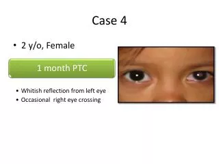
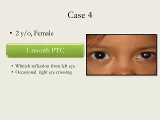
Case 4 • 2 y/o, Female
Clinical Impression RETINOBLASTOMA
What is the differential diagnosis? • Retrolentalfibroplasia • Persistence of the primary vitreous • Retinal dysplasia • Coats’ disease • Nematode endophthalmitis
RetrolentalFibroplasia • Retinopathy of Prematurity • Disturbance in the retinal vascularization in premature infants • May be acute or chronic • Usually bilateral, often asymmetric • Causative role of supplemental oxygen
RetrolentalFibroplasia • Major risk factors • Decreasing gestational age • Decreasing birth weight • Other associated risk factors • Acidosis • Apnea • PDA • Septicemia • Blood transfusions • Intraventricularhemorrhage
RetrolentalFibroplasiaClinical Manifestations Reference: Nelson Textbook of Pediatrics. 18th Edition. 2007.
RetrolentalFibroplasiaClinical Findings Reference: Nelson Textbook of Pediatrics. 18th Edition. 2007.
Stage 1 ROP Stage 2 ROP Stage 3 ROP
Stage 4 ROP Stage 5 ROP
RetrolentalFibroplasiaDiagnosis • Recommendations • Systematic serial ophthalmologic examinations of infants at risk • Infants weighing <1500g at birth • Infants born before 31 weeks AOG • Infants weighing >1500g at birth who have an unstable clinical course Reference: Nelson Textbook of Pediatrics. 18th Edition. 2007.
RetrolentalFibroplasiaDiagnosis • Recommendations • Initial examination should be performed at 4th to 6th week of chronological age • ROP is diagnosed most often at 32nd to 44th week after conception. • Screening should continue until the retina is fully vascularized, until the changes of ROP have undergone spontaneous resolution, or until appropriate treatment has been given. Reference: Nelson Textbook of Pediatrics. 18th Edition. 2007.
RetrolentalFibroplasiaTreatment • Early Treatment of Retinopathy of Prematurity Study • Recommends treatment for infants with Stage 2 disease with vascular tortuosity and engorgement • Laser – treatment modality of choice • Vitrectomy and lensectomy in cicatricial disease References: Nelson Textbook of Pediatrics. 18th Edition. 2007. Vaughan & Asbury’s General Ophthalmology 17th Edition. 2008.
Persistent hyperplastic primary vitreous (PHPV) • a congenital developmental anomaly of the eye resulting from failure of the embryological, primary vitreous and hyaloid vasculature to regress, whereby the eye is shorter, develops a cataract, and may present with whitening of the pupil • children are born with a hazy, scarred vitreous, which blocks light passing to the back of the eye, leading to blurred vision
The scarred vitreous is often stuck to the back of the lens and areas of the retina. This can damage these parts of the eye as well and lead to other eye conditions: • the lens may become hazy (cataract) • the retina may peel off the back of the eye (retinal detachment) • the pressure in the eye can rise (glaucoma) • the eye can be smaller than usual (microphthalmia)
Why are some children born with PHPV? • in children with PHPV, the first (primary) vitreous that grows in the eye fails to become clear • it grows too much (hyperplastic) and becomes hazy and scarred • Usually it would disappear and become clear but instead it stays (persists) • the etiology is unknown • most cases of PHPV occur by chance, although some cases are known to run in families • usually the condition only affects one eye
It can be present in three forms: • purely anterior (persistent tunica vasculosalentis and persistent posterior fetal fibrovascular sheath of the lens) • purely posterior (falciform retinal septum and ablatiofalcicormiscongentia) • combination of both
How is PHPV first suspected? • PHPV can present in different ways. These include: • White pupil (leucocoria) – MOST COMMON PRESENTATION • Squint (when both eyes don't appear to point in the same direction) • Painful red eye • Blurred vision • Nystagmus
Most children with PHPV in only one eye usually have good vision in the other. These children do not normally complain of reduced vision. • If both a child's eyes suffer from PHPV then they are much more likely to have serious visual impairment. They may only see bright lights and large shapes. This usually allows the child to get around but may not notice kerbs and smaller objects on the floor. They are likely to recognize faces from close up and be able to play with large toys.
Treatment depends on: • Whether both eyes are affected or not and • What other eye conditions develop • If only one eye is affected operations are avoided. The main aim is to try to preserve the eye. If the other eye has good vision it is often best not to try operations that are unlikely to offer an affected child any overall visual benefit. • If both eyes are affected operations can be done to try to improve vision. If an eye becomes red and painful due to high pressure operations may also be needed. An operation normally tries to take away the hazy vitreous and usually the lens as well. This would let light travel through to the back of the eye without being blocked. It also helps to reduce pressure in the eye.
Retinal dysplasia • usually a nonprogressive disease and can be caused by viral infections, drugs, vitamin A deficiency, or genetic defects • is characterized by folds or rosettes (round clumps) of the retinal tissue
Coats’ Disease exudative retinitis retinal telangiectasis • very rare congenital, nonhereditary eye disorder, causing full or partial blindness, characterized by abnormal development of blood vessels behind the retina • result from breakdown of the blood-retinal barrier in the endothelial cell, resulting in leakage of blood products containing cholesterol crystals and lipid-laden macrophages into the retina and subretinal space. Over time, the accumulation of this proteinaceousexudate thickens the retina, leading to massive, exudative retinal detachment. Patient with Coats' disease, showing conjunctival hyperemia, mild corneal edema, posterior synechiae and cataract.
Presentation • Unilateral • Predominantly in young males • Peak age of onset: 6-8 years of age • Results in a gradual loss of vision • Progresses slowly • At advanced stages, retinal detachment is likely to occur • Glaucoma,atrophy, and cataracts can develop secondary to Coats’ disease • Most common sign at presentation: leukocoria • Symptoms typically begin as blurred vision, usually pronounced when one eye is closed due to the unilateral nature of the disease • Flashes of light (photopsia and floaters) are common symptoms • One early warning sign of Coats’ disease is yellow-eye in flash photography • Coats’ disease itself is painless. Pain may occur if fluid is unable to drain from the eye properly, causing the internal pressure to swell, resulting in painful glaucoma.
Five Stages Based on the severity of the abnormalities that resulted from the vascular changes (1965, Gomez Morales): Stage 1: only focal exudates Stage 2: massive intraretinal exudation Stage 3: partial exudative retinal detachment Stage 4: total retinal detachment Stage 5: complications secondary to chronic retinal detachment (neovascular glaucoma) • Based selecting treatment and predicting the ocular and visual outcomes of the disease (Shields et al.): • Stage 1: retinal telangiectasia • Stage 2: telangiectasia and exudation • A: extrafoveal exudation • B:foveal exudation • Stage 3: exudative retinal detachment • A: subtotal detachment • 1: extrafoveal • 2: foveal • B: total retinal detachment • Stage 4: total retinal detachment and glaucoma • Stage 5: advanced end-stage disease
Diagnosis • Slit lamp biomicroscopy • normal findings of anterior segment • differentiation of Coats disease from congenital cataract and persistent hyperplastic primary vitreous • Funduscopic eye examination • retinal vessels in early Coats' disease appear tortuous and dilated, mainly confined to the peripheral and temporal portions of retina • moderate to severe Coat's disease, massive retinal detachment and hemorrhage from the abnormal • Fundus examination with indirect ophthalmoscopy and detailed, large fundus drawing, fundus photography and fluorescein angiography • differentiating Coats disease from retinoblastoma.
Diagnosis • Ultrasound • hyperechoic mass in the posterior vitreous without posterior acoustic shadowing; vitreous and subretinal hemorrhage • CT • globe appears hyperdense compared to normal vitreous due to the proteinaceousexudate, which may obliterate the vitreous space in advanced disease • anterior margin of the subretinalexudate enhances with contrast. • retina is fixed posteriorly at the optic disc, this enhancement has a V-shaped configuration • MRI • subretinalexudate shows high signal intensity on both T1- and T2-weighted images • exudate may appear heterogeneous if hemorrhage or fibrosis is present • subretinal space does not enhance with gadolinium contrast. • Mild to moderate linear enhancement may be seen between the exudate and the remaining vitreous. • exudate shows a large peak at 1-1.6 ppmonproton MR spectroscopy.
Diagnosis Computed Tomography image of a patient with Coats' disease, showing total exsudative retinal detachment in the right eye.
Diagnosis • Grossly • retinal detachment and yellowish subretinalexudate containing cholesterol crystals • Microscopically • wall of retinal vessels may be thickened in some cases, while in other cases the wall may be thinned with irregular dilatation of the lumen • subretinalexudate consists of cholesterol crystals, macrophages laden with cholesterol and pigment, erythrocytes, and hemosiderin • granulomatous reaction, induced by the exudate, may be seen with the retina
Diagnosis A case of Coats' disease, showing total retinal detachment with subretinalexudate containing cholesterol crystals and a fibrous nodule in the posterior pole. A case of Coats' disease, showing total exsudative retinal detachment, and subretinalexudate containing cholesterol crystals (H&E).
Treatment • Stage 1 disease (telangiectasia only) • either periodic observations or laser photocoagulation • high probability that the eye can be saved • visual prognosis is usually favorable • uncommon in a clinical practice, usually advanced at the time of diagnosis. • Stage 2 disease (telangiectasia and exudation) • laser photocoagulation or cryotherapy, depending on the extent of the disease and the preference of the ophthalmologist • exudation is limited to one quadrant or located nasally, a reasonably good visual outcome • stage 2A • visual prognosis is generally good, because the fovea is not involved by exudation. • stage 2B • visual prognosis is relatively good if the foveal exudation is not advanced • associated with a dense yellow gray nodule centered within the foveal exudation, the visual prognosis worsens
Treatment • Stage 3A disease (subretinal retinal detachment) • photocoagulation or cryotherapy • Even if the retinal detachment involves fovea, it will resolve when the telangiectasia is eradicated • subretinal fluid in the retinal detachment makes laser photocoagulation less effective than cryotherapy. • Stage 3B disease (total retinal detachment) • cryotherapy if the retinal detachment is shallow, but may require an attempt at surgical reattachment if the detachment is advanced and immediately posterior to the lens. • Stage 4 disease (total retinal detachment with glaucoma) • enucleation for the severe ocular pain • Stage 5 disease • patients have generally blind, but comfortable eye • require no aggressive treatment
Treatment Surgical treatment • Coats disease complicated by exudative retinal detachment • pars planavitrectomy to drain the subretinal fluid, thus allowing the treatment of pathologic vessels • removal of tractional vitreous membranes, mostly invisible, can be required to reattach the retina • removal of the epiretinal membrane and endocryocoagulation of the affected retina result in a complete resorption of subretinal fluid exudates
Nematode Endophthalmitis • Inflammatory condition of the intraocular cavities (aqueous or vitreous humor) usually caused by infection. • Noninfectious (sterile) endophthalmitis may result from various causes such as retained native lens material after an operation or from toxic agents • Two types of endophthalmitis are endogenous (metastatic) and exogenous • Endogenous endophthalmitis results from the hematogenous spread of organisms from a distant source of infection (endocarditis) • Exogenous endophthalmitis results from direct inoculation as a complication of ocular surgery, foreign bodies, and/or blunt or penetrating ocular trauma.
Presentation SYMPTOMS • visual loss • eye pain and irritation • Headache • Photophobia • ocular changes • intense ocular and periocular inflammation PHYSICAL EXAMINATION • Eyelid swelling and erythema • Hypopyon, Vitreitis, and Chemosis • Reduced or absent red reflex • Proptosis • Papillitis • Cotton-wool spots • Corneal edema and infection • White lesions in the choroid and retina • Chronic uveitis and vitreal mass and debris • Purulent discharge • Fever • Cells and flare in the anterior chamber on slit lamp examination .
Diagnosis • check of visual acuity • examine both eyes by slit lamp biomicroscopy • intraocular pressure • dilated funduscopy • ultrasonography if fundus not well visualized help determine if a retained intraocular foreign body is present, the density of the vitreitis, and if the retina is attached or not • routine cultures should include aerobic, anaerobic, and fungal cultures
Treatment • Once the diagnosis has been made, or strongly considered, prompt consultation to an ophthalmologist is needed. • Treatment depends on the underlying cause of endophthalmitis.
Onchocerciasis • nematode infection in humans that affect the eye • caused by O. volvulus. • Ocular changes • oncintraocularmicrofilariae • punctatekeratitis • sclerosingkeratitis • anterior uveitischorioretinitis • optic neuritis • optic atrophy • Glaucoma • river blindness. • Diagnostic tests • skin snip • Nodulectomy • slit-lamp examination, • Mazzotti test
Onchocerciasis • Goals of pharmacotherapy: eradicate the infestation, to reduce morbidity, and to prevent complications • Anthelmintic agents can be used • Parasite biochemical pathways are different from the human host; thus, toxicity is directed to the parasite, egg, or larvae. • Ivermectin (Mectizan) 150 mcg/kg PO as single dose; may repeat q6-12mo until asymptomatic can be used for Cutaneous larva migrans (onchocerciasis)
CBC, electrolyte determination, urinalysis, liver function test to exclude other conditions cofused with retinoblastoma • Aqueous humor enzyme level – aqueous humor lactate dehydrogenase (LDH) to serum LDH ratio • < 1.0 in patients with ocular disease other than retinoblastoma • > 1.0 – aqueous humor for eyes with retinoblastoma exhibits increased LDH activity
Cranial and orbital computerized tomography • Sensitive for diagnosis and detecting intraocular calcification • shows intraocular extent of the tumor even in the absence of calcification. • also invaluable in assessing the CNS anatomy, including the optic nerve, for possible extension of retinoblastoma.
Ultrasonography • distinguishing retinoblastomas from non-neoplastic conditions. • also useful in detecting calcifications. • MRI • estimating the degree of differentiation of retinoblastomas but is not as specific as computerized tomography because of its lack of sensitivity in detecting calcium. • identifying any associated hemorrhagic or exudative retinal detachment. • seen as a localized subretinal area of higher signal intensity compared to vitreous on both T1- and T2-weighted sequences. seen as an irregular mass lesion in the vitreous cavity with calcification. Presence of calcification is helpful in the diagnosis of retinoblastoma, but absence does not rule out retinoblastoma. On A-Scan there is irregular high reflectivity with distal shadowing. MRI T1 weight contrast enhanced image of retinoblastoma (Image from Abeloff, M.D., Clinical oncology. 3rd ed. 2004, Philadelphia, Pa.: Elsevier Churchill Livingstone. xxiv, 3205.)
X-ray studies: • In areas of the world where ultrasonography and computerized tomography are not available, x-ray studies may be the only means of identifying intraocular calcium in patients with opaque media.
GENETIC COUNSELING • Blood specimens should be taken not only from the patient but also from the parents and any siblings for DNA analysis • direct method aims to find the initial mutation that precipitated the development of the tumor; then, it is determined whether that mutation is in the germline of the affected patient. • Indirect methods can be used in cases where the initial mutation cannot be located or it is uncertain whether it exists.
Immunohistopathologic staining • to decide whether retinoblastomas come from a common progenitor cell capable of differentiation into either glial or neuronal cells or from neuron-committed cells. • show an S-antigen detected in well-differentiated retinoblastomas using immunoperoxidase staining of paraffin sections • Transmission electron microscopy • provided evidence of the presence of photoreceptor cell elements in retinoblastoma, and a strong evidence of retinoblastoma to human fetal retina
Patients noted to have presenting signs of retinoblastoma should undergo prompt office examination • Complete eye examination should be performed including an estimation of the patient's visual acuity for both eyes • A dilated fundus examination with indirect ophthalmoscopy should be completed since ancillary diagnostic studies play only a secondary role when the fundus can be visualized clearly
A bone marrow aspiration and biopsy could be performed as well as lumbar puncture with cytocentrifuge examination for tumor cells. These may prove useful in the early diagnosis of distant spread since the primary mode of spread of retinoblastoma is hematogenous to the bone marrow and back through the optic nerve into the cerebrospinal fluid (CSF)