Download
1 / 13
130 likes | 331 Views
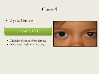
Case 4. 2 y/o, Female. Case 4. Case 4. Clinical Impression. RETINOBLASTOMA. What is the differential diagnosis?. p.209 (book) Retrolental fibroplasia Persistence of the primary vitreous Retinal dysplasia Coats’ disease Nematode endophthalmitis.

E N D
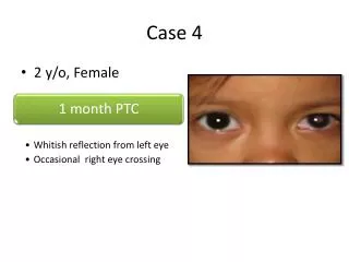
Case 4 • 2 y/o, Female
Clinical Impression RETINOBLASTOMA
What is the differential diagnosis? • p.209 (book) • Retrolentalfibroplasia • Persistence of the primary vitreous • Retinal dysplasia • Coats’ disease • Nematode endophthalmitis
a congenital developmental anomaly of the eye resulting from failure of the embryological, primary vitreous and hyaloid vasculature to regress, whereby the eye is shorter, develops a cataract, and may present with whitening of the pupil • children are born with a hazy, scarred vitreous, which blocks light passing to the back of the eye, leading to blurred vision
The scarred vitreous is often stuck to the back of the lens and areas of the retina. This can damage these parts of the eye as well and lead to other eye conditions: • the lens may become hazy (cataract) • the retina may peel off the back of the eye (retinal detachment) • the pressure in the eye can rise (glaucoma) • the eye can be smaller than usual (microphthalmia)
Why are some children born with PHPV? • in children with PHPV, the first (primary) vitreous that grows in the eye fails to become clear • it grows too much (hyperplastic) and becomes hazy and scarred • Usually it would disappear and become clear but instead it stays (persists) • the etiology is unknown • most cases of PHPV occur by chance, although some cases are known to run in families • usually the condition only affects one eye
It can be present in three forms: • purely anterior (persistent tunica vasculosalentis and persistent posterior fetal fibrovascular sheath of the lens) • purely posterior (falciform retinal septum and ablatiofalcicormiscongentia) • combination of both
How is PHPV first suspected? • PHPV can present in different ways. These include: • White pupil (leucocoria) – MOST COMMON PRESENTATION • Squint (when both eyes don't appear to point in the same direction) • Painful red eye • Blurred vision • Nystagmus
Most children with PHPV in only one eye usually have good vision in the other. These children do not normally complain of reduced vision. • If both a child's eyes suffer from PHPV then they are much more likely to have serious visual impairment. They may only see bright lights and large shapes. This usually allows the child to get around but may not notice kerbs and smaller objects on the floor. They are likely to recognize faces from close up and be able to play with large toys.
Treatment depends on: • Whether both eyes are affected or not and • What other eye conditions develop • If only one eye is affected operations are avoided. The main aim is to try to preserve the eye. If the other eye has good vision it is often best not to try operations that are unlikely to offer an affected child any overall visual benefit. • If both eyes are affected operations can be done to try to improve vision. If an eye becomes red and painful due to high pressure operations may also be needed. An operation normally tries to take away the hazy vitreous and usually the lens as well. This would let light travel through to the back of the eye without being blocked. It also helps to reduce pressure in the eye.
Retinal dysplasia • usually a nonprogressive disease and can be caused by viral infections, drugs, vitamin A deficiency, or genetic defects • is characterized by folds or rosettes (round clumps) of the retinal tissue