Cervical and Lumbar Evaluations
390 likes | 1.13k Views
Cervical and Lumbar Evaluations. Inservice by Brittany Annis Ithaca College. Spinal Evaluation. Upper Quarter: Cervical Spine-T4 Includes scapula/UE Lower Quarter: T4-Sacral spine Includes hip/LE.

Cervical and Lumbar Evaluations
E N D
Presentation Transcript
Cervical and Lumbar Evaluations Inservice by Brittany Annis Ithaca College
Spinal Evaluation • Upper Quarter: • Cervical Spine-T4 • Includes scapula/UE • Lower Quarter: • T4-Sacral spine • Includes hip/LE • “Most back disorders are not the result of a single traumatic injury. Rather, they are the result of the cumulative effects of poor posture, faulty body mechanics, stressful living and working habits, loss of strength and flexibility and a general decline in the level of physical fitness." Saunders 1993.
Spinal Evaluation • History • Observation/Posture • Examination: • AROM • PROM • Resisted isometrics • Peripheral joint scan • Myotomes • Special Tests • Reflexes/Dermatomes • Segmental motion/joint play • Palpation • Assessment • Plan
Normal Cervical Range • Flexion/Extension: • Suboccipital: 20-35 degrees • Mid-cervical: 30-35 degrees • Sidebending: • A/A: 20 degrees • Mid-Cervical: 25 degrees • Rotation: • Sub-Occipital: 35 degrees • Mid-Cervical: 45 degrees
Normal Thoracic Range • Flexion: 20-40 degrees • Extension: 15-30 degrees • Sidebending: 25-30 degrees • Rotation: 5-20 degrees
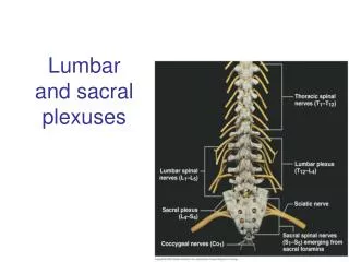
Normal Lumbar Ranges • Flexion (greatest at L4-L5): 40-60 degrees • Extension: 20-25 degrees • Sidebending (greatest at L3-L4) 15-35 degrees • Rotation (greatest at L4-S1): 5-20 degrees
Postural Assessment • Check anterior-posterior curves (kyphosis/lordosis) • Check lateral curves (scoliosis) • Check superior/inferior symmetry (ribs/skin folds)
Muscles prone to tightness(postural/static) • Sternocleidomastoid • Scalenes • Levator Scap • Pec major • Upper trapezius • Upper extremity flexors • Quadratus lumborum • Back extensors • Erector Spinae, Rotators, Multifidi • Hip flexor • Iliopsoas, tensor fasciae latae, rectus femoris • Lateral hip rotators (piriformis) • Proximal Hip adductors • Hamstrings • Foot plantarflexors • Gastroc/soleus, tibialis posterior
Muscles prone to weakness(dynamic) • Short neck flexors • Lower trap • Rhomboids • Serratus anterior • Subscapularis • Upper extremity extensors • Rectus abdominus • Internal/external obliques • Gluteus maximus/medius/minimus • Vastus medialis/lateralis • Tibialis anterior • Peroneous longus/brevis
Increased Kyphosis and Decreased Lordosis • Posterior pelvic tilt • Tight hip extensors • Weak hip flexors/ lower abdominals • Altered GH rhythm due to resting position of scapula: forward head and shoulders • Genu Recurvatum: increased stress on posterior knee and compression on anterior knee • Increased stress on anterior hip jt, and posterior thoracic spine • Shortening of posterior hip ligaments and anterior thoracic spine ligaments
Increased Lordosis: • Anterior pelvic tilt • Tight hip flexors/back extensors • Weak hip extensors/abdominals • Increased shear forces on lumbar vertebrae • Increased compression forces on lumbar facets • Increased stress on anterior spinal ligaments • Narrowing of lumbar intervertebral foramen
Movement Assessment • Upper Quarter • Scapulohumeral rhythm: assess glenohumeral flexion/abduction • Lower Quarter • Standing: single leg stance: stand on left leg, should feel erector spinae contract on right • Sitting: lateral weight shift to L buttock, assess curve of the spine and contraction of muscles
Lumbar AROM • Forward Flexion: • Does normal lordosis flatten (normal)? Round into kyphosis (hypermobility)? Stay in lordosis (hypomobile)? How much standing hip flexion? Is movement smooth throughout range? Pain/stiffness/other symptoms? • "torturous recovery" with active lesion in spinal muscles if painful to stand back up into extension, when mm are actively contracting; verses inert soft tissue like capsules or ligaments will be painful at the end range of motion, and will loosen up with repeated movement. • Drift to left or right instead of straight up when returning from forward flexion? If so, unilateral hypomobility/tightness of soft tissue likely exists. • Backbending • May be restricted by disc protrusion, soft tissue hypomobility, facet joint pain • Pain at end range indicated soft tissue hypomobility/facet; repetitive back bending with pain but not an increase in pain indicated stiffness
Lumbar AROM, con’t • Sidebending • Measure distance of fingers down lateral side of leg to floor; compare bilaterally • Look for gentle, continuous, smooth curve from lumbosacral joint to mid-thoracic area • If straight area, hypomobility is present • If sharp bending, hypermobility is present • Rotation • Arms across chest, assess spinous processes bilaterally
Lumbopelvic Rhythm • Interconnection of movement between spine and pelvis • Forward flexion: lumbar lordosis curve reverses from concave to slightly convex as the pelvis rotates about the hips. Equal balance between lumbar reversal and pelvic rotation. • Lack of equal motion: facet restriction, degenerative jt disease, tight hamstring muscles • Lumbar spine must fully reverse itself, and pelvis must rotate to full extent. • Hips and pelvis are posteriorly displaced in the horizontal plane to maintain the center of gravity over the feet, so the person doesn't fall forward. • Return from flexion: opposite occurs. Lumbar spine becomes concave, pelvis derotates, and shifts forward in horizontal plane. Same smoothness in both directions. • Restrictions in this motion can occur at the hips as well; if the patient feels restriction/pain at the hip, may be preventing full flexion
PROM • Passive: differentiates between contractile and non-contractile structures. If passive ROM is greater than AROM, contractile tissue is in part responsible for the pt's symptoms. If AROM is greater than PROM, pt likely cannot relax enough to move through motion. • Flexion: • Supine, hug knees to chest; repeat several times without letting go of the knees between repetitions. Assess whether pain increases or decreases. • Stretches lumbar muscles, but also compresses the disc, which may result in pain and peripheral symptoms. • Extension • Perform a prone press-up, with hands directly underneath shoulders. Repeat the movement several times to assess whether repetitions worsen or decrease symptoms. • Determine if a normal curve exists with increase in lordosis • Test hip IR, ER, extension, flexion and assess for provocation of symptoms
Segmental Motion • Segmental motion pinpoints exactly where the abnormal motion is occurring, whereas PROM gives the clinician only a generalized picture of joint mobility. • If the pt has full AROM/PROM, the segmental tests are still useful because abnormal joint mechanics may still be present. • One joint may be extremely stiff, but it is undetectable with AROM/PROM because other joints are mobile enough to compensate for it, which overall appears to be normal ROM. • Palpate to find tender spinous/transverse processes or interspinous spaces, then perform specific segmental mobility tests to identify differences in mobility between tender and non-tender segments. • If tender segments move more, they are hypermobile. If they move less, they are hypomobile.
Lumbar Segmental Motion • During forward flexion, while seated or in prayer position, assess the separation of spinous processes from vertebral levels and compare. • During prone press up into extension, assess the spinous processes moving together, and compare to other levels • Assess lumbar rotation when performing active rotation; palpate interspinous space as the patient actively rotates; compare adjacent segments. • Spring testing: Normalized anterior/posterior pressure through each segment, determine specific stuff/painful segments
Palpation • Palpate skin for tenderness, color, temperature, moisture, texture • Palpate ligaments: may be tender if sprained or inflamed; may be thickened if joint is hypomobile • Palpate bones to rule out dislocation/subluxation/ facet joint impingement • Palpate muscles for guarding/spasm; will feel tough/ropy
Palpation • Sacrotuberous ligament: from posterior sacrum to ischial tuberosities • If unusually tight, consider posterior innominate rotation, resulting in taught ligament • If unusually springy or unpalpable, consider anterior innominate rotation, increasing slack on ligament • Weight–shift test: stand on single leg, palpate lumbar paraspinals on contralateral side; switch to other side and compare. • Paraspinal muscles on stance foot should relax. If not, there is likely presence of muscle spasm/guarding
Palpation • Piriformis: • Patient lies prone, palpate halfway between the ischial tuberosity and greater trochanter; assess for soreness and difference from side to side • Iliopsoas: 3 places: • 1) Inside of the ilium, medial to the ASIS • 2) Anterior hip, lateral border of the femoral triangle • 3) Downward and medially, 2 inches lateral to umbilicus • Quadratus lumborum • Standing, sidebend and rotate to opposite side, palpate just lateral to paraspinal muscles
Flexibility • Important to note whether the range of motion at a joint is limited by intrinsic joint structures or by the muscles crossing the joint • Consider tonal/resistance changes, quality/quantity and range of movement • Muscle length testing allows clinician to evaluate muscle tone: hypertonic, tight, adaptive shortening • Consider pain behavior: referred and local pain throughout the range • Hypermobility= excessive elongation of the myofascial system, joint capsule laxity, or laxity of supporting ligaments.
Flexibility Testing: • Latissimus Dorsi: Hooklying, arms overhead, try to get arms flat on table. Look for lumbar extension or arms moving out of sagittal plane, representing tightness and resulting in increased lordosis • Hamstring (90/90): If hamstrings are lacking 20 degrees extension, they are considered tight and can affect lumbar lordosis • Rectus femoris/Iliopsoas: Decreased flexibility can pull pelvis into anterior pelvic tilt, which consequently increases lumbar lordosis • Pec Minor: In supine, asymmetrical shoulder height from table • Could also be tight joint capsule, differentiate by spring testing. Decreased AP joint motion over coracoid is pec minor tightness, whereas decreased AP joint motion over humeral head is glenohumeral tightness.
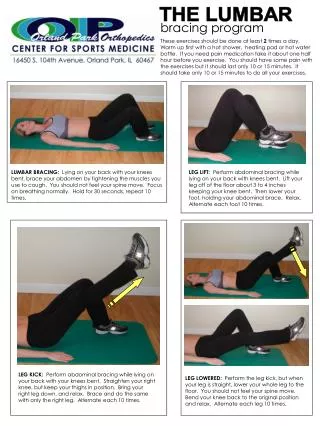
Strength test: • Abdominal strength test: • Patient in half-sit up position; must hold for over 10 seconds. If unable, abdominal muscles need strengthening • Lumbar paraspinals strength test: • Lying prone with arms overhead, lift chest off table. (If limited spine extension ROM, have upper body off end of table). • Hold position off the table for over 10 seconds. If unable, the pt needs aggressive strengthening. • Demo
Joint Mobility • Quality /quantity of motion; end feel • Normal joints are smooth, friction-free, with healthy amount of joint play at end-range • Pathologic joints have friction, joint sounds, and deviations from normal direction of motion; may feel restricted, blocked, painful, or abnormally loose • If joint dysfunction is present, there is almost always a loss of myofascial extensibility as a result. In order to effectively treat the joint hypomobility, the myofascial component needs to be released first! • Holistic approach, rather than a localistic approach
Type I vs Type II Restrictions • Type I: multisegmental restrictions. Are "accommodating, adapting, or neutral" from anatomic/functional asymmetries, SI dysfunctions, posture dysfunctions, etc (scoliosis) Most likely limited in sidebending • Type II: unisegmental: "non-adapting, non-accommodating" lesions, usually traumatic in nature (fall, slip) Will likely restrict the segment above and below it. Flexion/extension most affected components. • “Most back disorders are not the result of a single traumatic injury. Rather, they are the result of the cumulative effects of poor posture, faulty body mechanics, stressful living and working habits, loss of strength and flexibility and a general decline in the level of physical fitness." principles of spinal evaluation and treatment book
Special Tests: • Vertebral Artery: Pt lays supine; PT extends, rotates, and laterally flexes neck to each side. Hold position shortly and observe for 5 D's (dizziness, diplopia, dropsy, dysarthria, dysphagia), signifying partial or complete occlusion of vertebral aa on that side. • Slump: Seated at edge of table with slumped spine and flexed neck. Examiner passively extends knee, then dorsiflexes foot. Positive sign pain/discomfort or neurological symptoms down tested LE; extend neck and if symptoms disappear, the test is positive. • Faber’s: Patient is supine, passively flex, abduct, and externally rotate the LE so that the foot rests on the opposite knee. Examiner applies pressure on the involved knee towards the table. Positive test is tightness/discomfort at hip. • Mod Thomas: Patient is supine, with one knee to chest and tested LE out straight. A positive test reveals hip flexor tightness on the extended leg, which will be lifted off of the table. • Differs from Thomas test, which tests for rectus femoris tightness and has patient’s tested LE extended with knee bent over edge of table.
Dysfunction • Dysfunction is sub-grouped into classes: • -Class 1: muscular • -Class 2: shortening of ligaments • -Class 3: true articular hypomobility • -The major area of hypomobility is the driving force that throws off the rest of the body's musculoskeletal system- • *need to treat the primary area* before treating symptoms. Primary area may be asymptomatic and away from area of main complaint.
McKenzie’s Three Syndromes • Postural Syndrome • Common in <30 years old • Symptoms appear localized around the spine and does not refer distally • Pain brought on by mechanical stress of healthy tissue that has been loaded statically over long periods of time; pain is resolved when the tissue is unloaded • There is no acute inflammation, so the pain is not constant or induced by movement • Difficult to diagnose by examination due to no underlying tissue pathology; only positive finding is pain with consistent loading at end-range, statically. • Treated by correcting the faulty postural alignment when pain occurs, such as lying, standing, walking, or sitting. Ergonomic/ living space assessment • If not treated properly, can result in dysfunction syndrome.
McKenzie, con’t • Dysfunction Syndrome • Stems from an untreated postural syndrome • Pathological shortening/weakening of normal connective tissues, resulting in faulty alignment and musculoskeletal imbalances • Detrimental to work and/or home life • Symptoms appear at end range, not during movement. Pain is intermittent (like postural syndrome) but normal soft tissues are abnormally tight. • Pain does not refer distally (unless nerve root is involved). • Pain occurs immediately when shortened tissues are overstretched. • Must be treated properly to avoid worsened pathology, or derangement syndrome.
McKenzie, Con’t • Derangement Syndrome • Can involve neurological signs/symptoms that can refer distally • Pain is commonly severe and disabling, usually occurring during movement. • History of poor posture • Disc material is displaced, based on an off-centered loading of the intervertebral disc.
Cleland, JA, Markowski, AM, Childs, JD. Current Concepts of Orthopedic Physical Therapy: The Cervical Spine. 2nd ed. 2006. • Konin, JG, Wilksten, DL, Isear, JA. Special Tests for Orthopedic Examination. NJ:Slack Incorporated. 1997. • Saunders HD, Saunders R. Evaluation, Treatment and Prevention of Musculoskeletal Disorders. Volume 1: Spine. 3rd Ed. Chaska: The Saunders Group; 1993. • Makofsky, HW. Spinal Manual therapy: An Introduction to Soft Tissue Mobilization, Spinal Manipulation, Therapeutic and Home Exercises. Thorofare, NJ:SLACK Incorporated. 2003. • Magee, DJ. Orthopedic Physical Assessment. 2nd ed. Philadelphia:W.B. Saunders Groups. 1992.