Download
1 / 33
400 likes | 737 Views
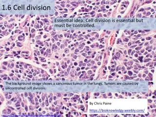
1.6 Cell division. Essential idea: Cell division is essential but must be controlled. The background image shows a cancerous tumor in the lungs. Tumors are caused by uncontrolled cell division. By Chris Paine https :// bioknowledgy.weebly.com /.

E N D
1.6 Cell division Essential idea: Cell division is essential but must be controlled. The background image shows a cancerous tumor in the lungs. Tumors are caused by uncontrolled cell division. By Chris Paine https://bioknowledgy.weebly.com/ http://www.flickr.com/photos/pulmonary_pathology/4388301142/
In summary any time new cells are required, mitosis is required: http://www.haroldsmithlab.com/images/pg_HeLa_cell_division.jpg
The cell cycle is the series of events through which cells pass to divide and create two identical daughter cells. http://upload.wikimedia.org/wikipedia/commons/thumb/7/71/Diagram_of_mitosis.svg/800px-Diagram_of_mitosis.svg.png
1.6.U4 Interphase is a very active phase of the cell cycle with many processes occurring in the nucleus and cytoplasm. Interphase consists of the parts of the cell cycle that don’t involve cell division. • G1 (Gap 1) • Increase the volume of cytoplasm • Organelles produced • Proteins synthesised • S (Synthesis) • DNA replicated • G2 (Gap 2) • Increase the volume of cytoplasm • Organelles produced • Proteins synthesised http://gardeningstudio.com/wp-content/uploads/the-cell-cycle-diagram-318.jpg
1.6.U4 Interphase is a very active phase of the cell cycle with many processes occurring in the nucleus and cytoplasm. Interphase Cells spend the majority of their time in interphase. It is a very active phase of the cycle. This when the cell carries out it’s normal functions Metabolic reactions (e.g. respiration to produce ATP) are necessary for the life of the cell Protein synthesis - proteins and enzymes are necessary to allow cell grow Organelles numbers are increased to first support the enlarged cell DNA is replicatedto ensure a second copy is available to enable mitosis Mr P O D http://botit.botany.wisc.edu/Resources/Botany/Mitosis/Allium/Various%20views/Interphase%20prophase.JPG
1.6.U1 Mitosis is division of the nucleus into two genetically identical daughter nuclei. Get the terminology right centrioles organise spindle microtubules centromereis the part of a chromosome that links sister chromatids Spindle microtubules (also referred to as spindle fibres) In animal cells two centrioles are held by a protein mass referred to as a centrosome Sister chromatidsare duplicated chromosomes attached by a centromere It is easy to misuse the terms chromatid and chromosome. It is even easier to confuse the terms centromere, centriole and centrosome due to their similar spelling. Keep the terms clear in your mind to avoid losing marks. After anaphase when the sister chromatids separate they should then be referred to as chromosomes http://commons.wikimedia.org/wiki/File:Chromosome.svg http://commons.wikimedia.org/wiki/Mitosis#mediaviewer/File:Mitosis_cells_sequence.svg
1.6.U1 Mitosis is division of the nucleus into two genetically identical daughter nuclei. Use the animated tutorials to learn about mitosis http://www.johnkyrk.com/mitosis.html http://highered.mheducation.com/sites/0072495855/student_view0/chapter2/animation__mitosis_and_cytokinesis.html http://outreach.mcb.harvard.edu/animations/cellcycle.swf http://www.sumanasinc.com/webcontent/animations/content/mitosis.html
1.6.U1 Mitosis is division of the nucleus into two genetically identical daughter nuclei. Prophase DNA supercoils* chromatin condenses and becomes sister chromatids, which are visible under a light microscope The centrosomes move to opposite poles of the cell and spindle fibres begin to form between them The nuclear membrane is broken down and disappears http://www.microscopy-uk.org.uk/mag/artnov04macro/jronionroot.html *supercoling is dealt with in more detail by 1.6.U2 http://commons.wikimedia.org/wiki/Mitosis#mediaviewer/File:Mitosis_cells_sequence.svg
1.6.U1 Mitosis is division of the nucleus into two genetically identical daughter nuclei. Metaphase • Spindle fibres from each of the two centrosomes attach to the centromere of each pair of sister chromatids • Contraction of the microtubule spindle fibres cause the sister chromatids to line up along the centre of the cell. http://www.microscopy-uk.org.uk/mag/artnov04macro/jronionroot.html http://commons.wikimedia.org/wiki/Mitosis#mediaviewer/File:Mitosis_cells_sequence.svg
1.6.U1 Mitosis is division of the nucleus into two genetically identical daughter nuclei. Continued contraction of the microtubule spindle fibres cause the separation of the sister chromatids The chromatids are now referred to as chromosomes Anaphase Chromosomes move to the opposite poles of the cell http://www.microscopy-uk.org.uk/mag/artnov04macro/jronionroot.html http://commons.wikimedia.org/wiki/Mitosis#mediaviewer/File:Mitosis_cells_sequence.svg
1.6.U1 Mitosis is division of the nucleus into two genetically identical daughter nuclei. Telophase The chromosomes uncoil de-condense to chromatin (and are no longer visible under a light microscope). Chromosomes arrive at the poles. Microtubule spindle fibers disappear New nuclear membranes reform around each set of chromosomes Now cytokinesis begins! http://www.microscopy-uk.org.uk/mag/artnov04macro/jronionroot.html http://commons.wikimedia.org/wiki/Mitosis#mediaviewer/File:Mitosis_cells_sequence.svg
1.6.U1 Mitosis is division of the nucleus into two genetically identical daughter nuclei. Prophase Metaphase Anaphase Telophase http://www.flickr.com/photos/chuckp/252924532/
1.6.U1 Mitosis is division of the nucleus into two genetically identical daughter nuclei. People Meet And Talk http://www.flickr.com/photos/chuckp/252924532/
1.6.U3 Cytokinesis occurs after mitosis and is different in plant and animal cells. • Urrrmm,we have only divided the nucleus … what about the rest of the cell? http://www.haroldsmithlab.com/images/pg_HeLa_cell_division.jpg
1.6.U3 Cytokinesis occurs after mitosis and is different in plant and animal cells. • mitosis is the division of the nucleus whereas • cytokinesisis the division of the cytoplasm • and hence the cell The division of the cell into two daughter cells (cytokinesis) occurs concurrently with telophase. Though mitosis is similar for animal and plant cells cytokinesis is very different. http://glencoe.mheducation.com/sites/9834092339/student_view0/chapter10/animation_-_cytokinesis.html http://www.haroldsmithlab.com/images/pg_HeLa_cell_division.jpg http://wwwprod.biochem.wisc.edu/biochem/faculty/bednarek/images/figure_color.gif http://commons.wikimedia.org/wiki/Mitosis#mediaviewer/File:Mitosis_cells_sequence.svg
1.6.U3 Cytokinesis occurs after mitosis and is different in plant and animal cells. http://www.haroldsmithlab.com/images/pg_HeLa_cell_division.jpg http://upload.wikimedia.org/wikibooks/en/thumb/9/98/Cyto.png/800px-Cyto.png
1.6.S1 Identification of phases of mitosis in cells viewed with a microscope or in a micrograph.1.6.S2 Determination of a mitotic index from a micrograph. http://www.nuffieldfoundation.org/practical-biology/investigating-mitosis-allium-root-tip-squash A very good, well explained lab outline for creating slides and calculating the mitotic index. An excellent online alternative if resources don’t permit students to create and view their own slides http://www.biology.arizona.edu/cell_bio/activities/cell_cycle/cell_cycle.html
1.6.U2 Chromosomes condense by supercoiling during mitosis. Why supercoil chromosomes? • Human cells are on average 10μm in diameter and the nucelus within each is less than 5 μm in diameter. • Human chromosomes are 15mm to 85mm (15,000μm to 85,000 μm) in length. • Chromosomes need to be stored compactly to fit within the nuclei of cells. • This problem becomes more acute during mitosis when chromosomes need to be short and compact enough that they can be separated and moved to each end of the cell. Chromatin fibres http://www.genome.gov/dmd/img.cfm?node=Photos/Graphics&id=85282
1.6.U2 Chromosomes condense by supercoiling during mitosis. How are chromosomes supercoiled? • Strain is placed on a DNA helix by overwinding or underwinding of the helix • This causes the DNA molecule to coil back on itself becoming shorter and wider • n.b.in eukaryotes proteins called histones aid the process Try it yourself http://vanat.cvm.umn.edu/mMeiosis/images/chromosome-X.jpg http://www.maths.uq.edu.au/~infinity/Infinity7/images/supercoiling.gif
1.6.U5 Cyclins are involved in the control of the cell cycle. Cyclinsare a family of proteins that control the progression of cells through the cell cycle 1 • Cells cannot progress to the next stage of the cell cycle unless the specific cyclin reaches it threshold. 2 • Cyclins bind to enzymes called cyclin-dependent kinases 3 • These kinases then become active and attach phosphate groups to other proteins in the cell. 4 • The attachment of phosphate triggers the other proteins to become active and carry out tasks (specific to one of the phases of the cell cycle). http://upload.wikimedia.org/wikipedia/commons/thumb/9/99/Protein_CCNE1_PDB_1w98.png/800px-Protein_CCNE1_PDB_1w98.png
1.6.U5 Cyclins are involved in the control of the cell cycle. Progression through parts of the cell cycle are affected in various ways by specific cyclins • prepares the cell for DNA replication in S phase. activates DNA replication inside the nucleus in S phase. • promotes the assembly of the mitotic spindle and other tasks in the cytoplasm to prepare for mitosis. Triggers cells to move from G0 to G1 and from G1 into S phase. http://upload.wikimedia.org/wikipedia/commons/thumb/9/99/Protein_CCNE1_PDB_1w98.png/800px-Protein_CCNE1_PDB_1w98.png
1.6.U6 Mutagens, oncogenes and metastasis are involved in the development of primary and secondary tumours. • Tumours are abnormal growth of tissue that develop at any stage of life in any part of the body. A cancer is a malignant tumour and is named after the part of the body where the cancer (primary tumour) first develops. Use the links to find out: • most common types of cancer • what causes cancer and associated risk factors • how cancer can be treated http://youtu.be/8BJ8_5Gyhg8 http://www.e-learningforkids.org/health/lesson/cancer/ What causes cancer? http://www.cancerresearchuk.org/cancer-info/cancerandresearch/all-about-cancer/what-is-cancer/ http://www.cancer.gov/cancertopics/types/commoncancers
1.6.U6 Mutagens, oncogenes and metastasis are involved in the development of primary and secondary tumours. • A mutation is a change in an organisms genetic code. A mutation/change in the base sequence of a certain genes can result in cancer. • Mutagens are agents that cause gene mutations. Not all mutations result in cancers, but anything that causes a mutation has the potential to cause a cancer. • Mutagens can be: • chemicals that cause mutations are referred to as carcinogens • high energy radiation such as X-rays • short-wave ultraviolet light • Someviruses http://en.wikipedia.org/wiki/Oncogene#mediaviewer/File:Oncogenes_illustration.jpg
1.6.U6 Mutagens, oncogenes and metastasis are involved in the development of primary and secondary tumours. If a mutation occurs in an oncogenesit can become cancerous. In normal cells oncogenes control of the cell cycle and cell division. mutation in a oncogene malfunction in the control of the cell cycle uncontrolled cell division tumour formation http://en.wikipedia.org/wiki/Oncogene#mediaviewer/File:Oncogenes_illustration.jpg
1.6.U6 Mutagens, oncogenes and metastasis are involved in the development of primary and secondary tumours. Several mutations must occur in the same cell for it to become a tumour causing cell. The probability of this happening in a single cell is extremely small. • Factors (other than exposure to mutagens) that increase the probability of tumour development include: • The vast number of cells in a human body – the greater the number of cells the greater the chance of a mutation. • The longer a life span the greater the chance of a mutation. http://en.wikipedia.org/wiki/Oncogene#mediaviewer/File:Oncogenes_illustration.jpg
1.6.U6 Mutagens, oncogenes and metastasis are involved in the development of primary and secondary tumours. The development of a primary tumours (cancers) have been outlined. Below is how a primary tumor can become a secondary tumour. • A primary tumor is a malignant tumor growing at the site where the abnormal growth first occurred. • Cancerous cells can detach from the primary tumour. • Metastasis is the movement of cells from a primary tumour to set up secondary tumours in other parts of the body. • Some cancerous cells gain the ability to penetrate the walls of lymph or blood vessels and hence circulate around the body • The circulating cancerous cells invade tissues at a different locations and develop, by uncontrolled cell division, into a secondary tumours. http://en.wikipedia.org/wiki/Neoplasm#mediaviewer/File:Colon_cancer_2.jpg
1.6.A1 The correlation between smoking and incidence of cancers. • There are many other similar surveys in different countries, with different demographics that show similar results. Along with lung cancer, cancers of mouth and throat are very common as these areas are in direct contact with the smoke too. It might surprise you that the following cancers are also more common in smokers: • Head and neck • Bladder • Kidneys • Breast • Pancreas • Colon Describe the relationship shown. What type of correlation is shown How strong is the correlation? Justify your answer by discussing the evidence. The correlation shown here is lagged. A lag is a time gap between the factors. Estimate the size of the lag between cigarette consumption and lung cancer death. http://en.wikipedia.org/wiki/File:Smoking_lung_cancer.png
1.6.A1 The correlation between smoking and incidence of cancers. • There are many other similar surveys in different countries, with different demographics that show similar results. Along with lung cancer, cancers of mouth and throat are very common as these areas are in direct contact with the smoke too. It might surprise you that the following cancers are also more common in smokers: • Head and neck • Bladder • Kidneys • Breast • Pancreas • Colon • Correlation ≠ causation, however laboratory investigations have found: • more then 20 chemicals found in tobacco have caused cancers in laboratory animals and/or humans • More than 40 other chemicals found in tobacco have been identified as carcinogens Describe the relationship shown. What type of correlation is shown How strong is the correlation? Justify your answer by discussing the evidence. The correlation shown here is lagged. A lag is a time gap between the factors. Estimate the size of the lag between cigarette consumption and lung cancer death. http://en.wikipedia.org/wiki/File:Smoking_lung_cancer.png
Bibliography / Acknowledgments Jason de Nys