Download
1 / 1
70 likes | 531 Views
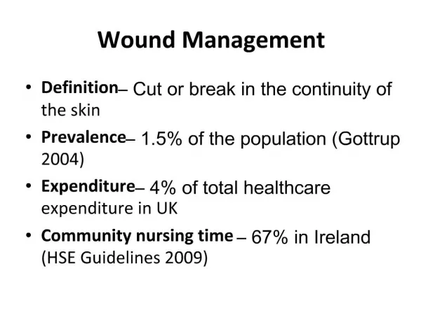
Standardization of Wound Photography Using the Wound Electronic Medical Record. USING WOUND PHOTOGRAPHY TO MEASURE HEALING/NONHEALING OVER TIME. A). B) . Day 0. Chandrasekaran E, Golinko M, Rennert R, Kaplan D, Brem H.

E N D
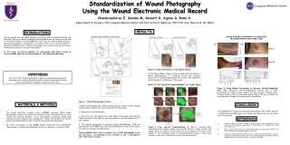
Standardization of Wound Photography Using the Wound Electronic Medical Record USING WOUND PHOTOGRAPHY TO MEASURE HEALING/NONHEALING OVER TIME A) B) Day 0 Chandrasekaran E, Golinko M, Rennert R, Kaplan D,Brem H. Department of Surgery, NYU Langone Medical Center and NYU Schol of Medicine, 530 First Ave, New York, NY 10016 Day 32 INTRODUCTION RESULTS Chronic wounds are a worldwide epidemic associated with increased morbidity and mortality. Objective wound evaluation and documentation are integral to the use of electronic medical records (EMR’s) to facilitate an evidence−based practice. Wound photography is recognized for its utility in the care of chronic wounds.1,2,3 Wounds calculated from a digital photograph are a quicker and more accurate method than contract tracing of the wound.1,2,3 In this study, we define guidelines for photography and digital tracing of chronic wounds based on an understanding of the underlying physiology. Day 0 Day 7 Day 14 Day 21 Day 28 Day 0 Day 14 Day 27 Day 41 HYPOTHESIS The use of the following guidelines to objective wound photography will decrease inter-observer variability and facilitate a consistent accurate clinical assessment of changes in wound area over time. Figure 2. Proper Orientation of the Digital Camera As the figure shows, changes in camera angle cause variability in wound measurements. To minimize errors and maintain objectivity of wound measurements, the camera lens should be oriented parallel to the plane of the wound. WHAT IS THE ACCURATE MEASUREMENT OF ULCER AREA? Figure 4. Using Wound Photography to Measure Healing/Nonhealing Over Time. Objective wound photography enables one to track progression of healing ulcers (Figure 4a) and calculate healing rates. Tracking healing rates of nonhealing ulcers (Figure 4b) can be helpful in deciding when a change of treatment plan is necessary. Figure 1. Wound Photography Protocol 1. Remove all dressings and remove as much necrotic tissue, slough, packing material and/or debris that can be tolerated by the patient. 2. Cleanse the wound with saline. 3. Place the ruler flat against the distal edge of the wound and hold the camera parallel to the plane of the wound with an optimal distance of 0.3 and 1 meters. 4. The digital photograph is measured using WoundImager 2.0® and entered into the WEMR along with wound dimensions, along with presence of cellulitis, drainage and undermining. These guidelines were used to capture over 14,000 photographs in the last four years, corresponding to the treatment of over 1,100 patients and 3,400 chronic wounds. MATERIALS & METHODS CONCLUSION The guidelines for wound documentation developed at this center provide a standardized methodology to accurately and objectively record wound area through time. The wound electronic medical record (WEMR) combines digital wound photography, recent area trends and relevant clinical information into an easily viewed and queried database. Strict photographic guidelines based upon individual wound etiologies were developed to reduce the variability of chronic wound area measurements and to facilitate informed clinical decisions. For more information on the WEMR, please attend the oral presentation “Development of a Relational Databank for Chronic Wounds,” on June 6, 2008 REFERENCES 1.) Samad A, Hayes S, French L, Dodds S. Digital imaging versus conventional contact tracing for the objective measurement of venous leg ulcers. J Wound Care. 2002 Apr;11(4):137-40. 2.) Ahn C, Salcido RS. Advances in wound photography and assessment methods. Adv Skin Wound Care. 2008 Feb;21(2):85-93; quiz 94-5. 3.) Golinko M, Chandrasekaran E, Cox D, Brem H. A novel relational database for clinical outcomes in patients with diabetic foot ulcers: The Wound Electronic Medical Record (WEMR). American College of Surgeons, Surgical Forum 2008. Figure 3. Ulcer Specific Troubleshooting. A.Callus is included when calculating wound length, width, and area. B. Necrotic tissue, eschar and slough, which are likely to be debrided, are still included in the area of the wound. C. All contiguous areas of non-epithelialized tissue in the same anatomic location are included in a single area measurement.