Download
1 / 59
590 likes | 712 Views
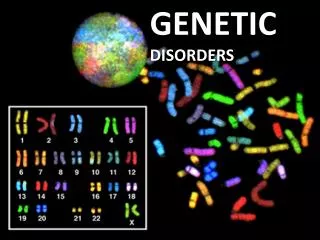
Genetic Disorders. INTRODUCTION: DEFINITION OF TERMS. CHROMOSOMES- cellular structures where genes are located GENES- basic units of heredity carry information necessary to determine specific biologic structures & functions ex. ABO Ag in RBC membrane coded by chromosome 9

E N D
INTRODUCTION:DEFINITION OF TERMS • CHROMOSOMES- cellular structures where genes are located • GENES- basic units of heredity carry information necessary to determine specific biologic structures & functions • ex. ABO Ag in RBC membrane coded by chromosome 9 • LOCUS- position in the chr where particular gene is located; all gene loci occur in pairs except X & Y genes
INTRODUCTION:DEFINITION OF TERMS • ALLELES- alternative genes in a single locus • ex. Kell blood group system • alleles K & k • KK- homozygous Kk- heterozygous • HOMOZYGOUS GENES- gene pair that are alike • HETEROZYGOUS GENES- gene pair that are not alike • GENOTYPE- actual gene composition that make the trait • PHENOTYPE- manifestation of the structure/ form produced by the genes
INTRODUCTION:DEFINITION OF TERMS • DOMINANT GENES- genes that are always expressed in the phenotype whether homozygous or heterozygous • RECESSIVE (AMORPH) GENES- genes that are masked if paired w/ a dominant gene, thereby only expressed when paired w/ another recessive gene
INTRODUCTION:DEFINITION OF TERMS • EUPLOIDY- multiples of the haploid # that is considered normal • HAPLOID (N)= 23- OCCURS IN MEIOSIS • DIPLOID (2N)= 46- OCCURS IN MITOSIS • ANEUPLOID- not exact multiples of the haploid #; only 1 pair of chr involved, therefore, germ cells have 2 copies of the same chr or lack the affected chr entirely • HYPODIPLOID 2N- 1, -2, ETC. (MONOSOMY) • HYPERDIPLOID 2N+ 1, +2, ETC. (TRISOMY)
INTRODUCTION:DEFINITION OF TERMS • POLYPLOID- multiples of haploid #; entire set of chrs fail to divide & all the chrs are segregated in a single daughter cell • TRIPLOID (3N)= 69 • TETRAPLOID (4N)= 92
Congenital Disorders • Non Genetic: • Developmental defects – Malformations • Genetic Disorders • Chromosomal • Gene - Mendelian • Multifactorial
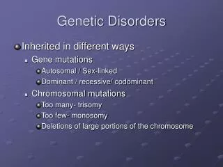
Mutations: • Genome: whole set – Polyploidy 4n, 8n etc. • Chromosomal: change in chromosome • Number: Trisomy, monosomy • Structure: Deletion, Translocation etc. • Gene: Submicroscopic • Point mutation – single base sequence • Deletions • Insertions
Cytogenetic Abnormalities: • Abnormal # of chrs: • Non-disjunction - Down’s Syndrome • Anaphase lag - Turner’s xxx • Abnormal Structure: (normal #) • Deletion - 5q- Cri - du - chat syndrome • Inversion - • Translocation - Ph Chromosome - t(9:22) CML,
GENETIC PATHOLOGY: • DEFINITION: Abnormalities or disease states that may or may not be congenital, transmitted by genes or chromosomal aberrations, that may be heritable (familial) or mutational • If mutational, may give the following outcomes: • Heritable • Disappear • Lethal • Sterility • Malignancy
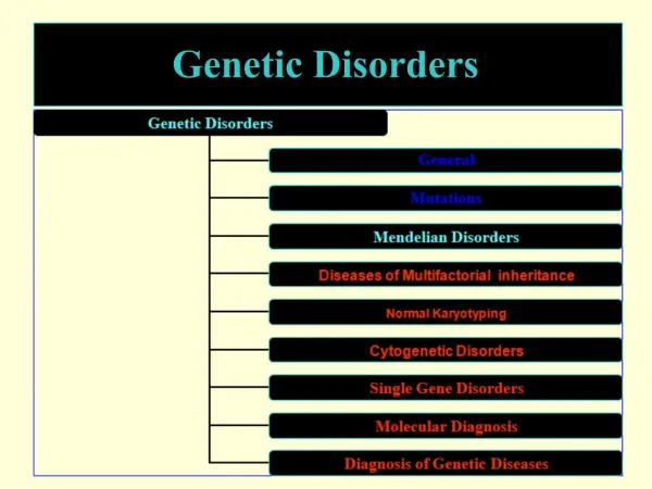
CATEGORIES: • CHROMOSOMAL ABNORMALITIES/ MUTATIONS • GENE ABNORMALITIES/ MUTATIONS • POLYGENIC/ MULTIFACTORIAL ABNORMALITIES
I. CHROMOSOMAL ABNORMALITIES/ MUTATIONS GENERAL CONCEPTS: • Children born to older women show more chromosomal aberrations than children born to younger women • Most major chromosomal abnormalities are incompatible w/ life • Detectable by karyotyping (chromosomal analysis) w/ or w/o banding techniques (use of stains)
I. CHROMOSOMAL ABNORMALITIES/ MUTATIONS::: TYPES: • NONDISJUNCTION (Chromosomal numerical aberration)- failure of chrs to sort themselves in equal #s into daughter cells • SUBTYPES: • POLYPLOIDY- see previous definition • ANEUPLOIDY- see previous definition • MOSSAICISM/ MYXOPLOIDY
I. ANEUPLOIDY: TRISOMY • TRISOMY- presence of 3 homologous chromosome in a cell • AUTOSOMAL TRISOMY- viable throughout pregnancy, even live born but die soon after birth except Down's syndrome • TRISOMY 21- DOWN'S SYNDROME • TRISOMY 18- EDWARD'S SYNDROME • TRISOMY 13- PATAU'S SYNDROME
I. ANEUPLOIDY: TRISOMY • SEX CHR. TRISOMY- abnormal development but non lethal; # of X chr. is directly proportional to mental retardation while number of Y chr. is directly proportional to aggressive behavior • TRIPLE X
I. ANEUPLOIDY: MONOSOMY • MONOSOMY- absence of one of a pair of homologous chr • AUTOSOMAL MONOSOMY- IUFD is the usual outcome • SEX CHR. MONOSOMY- compatible w/ life only if the conserved chr is an X, if not it will be less viable • TURNER'S SYNDROME- 45, XO
I. ANEUPLOIDY: MOSSAICISM/ MYXOPLOIDY • MOSSAICISM/ MYXOPLOIDY- nondisjunction at a later cell division resulting to population of normal & trisomic or monosomic cells coexisting in an individual • AUTOSOMAL MOSSAICISM- rare & lethal • SEX CHR. MOSSAICISM- common • GONADAL DYSGENESIS- TURNER'S SYNDROME 45, XO • KLINEFELTER'S SYNDROME 47 XXY
I. CHROMOSOMAL ABNORMALITIES/ MUTATIONS::: TYPES: I. MORPHOLOGIC/ STRUCTURAL SUBTYPES: • DELETION- loss of chromosomal material following a break in the chr arm or partial monosomy • CRI DU CHAT- partial monosomy of p5 • RETINOBLASTOMA- q13 • WILM'S TUMOR ANIRIDIA SYNDROME- p11
I. MORPHOLOGIC/ STRUCTURAL SUBTYPES: • TRANSLOCATION- transfer of segment of chromosomal material to another chromosome leading to imbalance of material in each daughter cell between non homologous chr • RECIPROCAL- acentric segments of chr exchanged for similar segment from a heterologous chr; use banding techniques for detection • ROBERTSONIAN (CENTRIC FUSION)- 2 acrocentric chr broken near centromere, exchange 2 arms and form new large metacentric chr and a small fragment, devoid of centromere & lost during subsequent division
I. MORPHOLOGIC/ STRUCTURAL SUBTYPES: • TRANSLOCATION • BALANCED- transfer w/ no loss of genetic material; individuals are normal except for infertility & if fertile, have a high risk of having malformed offspring • UNBALANCED- transmitted in the haploid gamete & paired w/ a new set of genes from the other parent • MALIGNANT LYMPHOMA- between 8 & 14 • LEUKEMIAS- between 22 & 9 • DOWN'S SYNDROME- chr 21
I. MORPHOLOGIC/ STRUCTURAL SUBTYPES: • TRANSLOCATION • ISOCHROMOSOMAL- faulty division of centromere at the transverse plane of the long axis w/ formation of a pair of isochromosome (one short arm & one long arm) • TURNER'S SYNDROME
I. MORPHOLOGIC/ STRUCTURAL SUBTYPES: • INVERSION- break of a chr at 2 points, followed by inversion of the intermediate segments & reunion results in the formation of a chr w/ rearranged distribution of genes • PERICENTRIC- rotation occurs around the centromere • PARACENTRIC- rotation occurs only on the acentric portion of the arm
I. MORPHOLOGIC/ STRUCTURAL SUBTYPES: • RING CHROMOSOME- break in the telomeric ends of the chr followed by deletion of the broken acentric segments & end to end fusion of the remaining portion
II. GENE ABNORMALITIES/ MUTATIONS GENERAL CONCEPTS: • Single gene defect detectable in the phenotype • Modified by penetrance, expressivity & whether defect is dominant, intermediate, recessive or X linked • Dominant pattern of inheritance usually due to alteration of aa sequence in the gene • Recessive pattern of inheritance (inborn errors of metabolism) usually is due to manufacture of abnormal enzymes or enzyme deficiencies • Follows Mendelian patterns of inheritance
PATTERNS OF INHERITANCE: AUTOSOMAL DOMINANT • Autosome- gene location • Gene expression- both homozygous & heterozygous state • Transmission of traits in every generation unless Low penetrance or modified by gene mutations • Unaffected family members do not transmit trait to offspring; affected family members usually heterozygous & transmit trait to only half of the offspring • M = F
PATTERNS OF INHERITANCE: AUTOSOMAL DOMINANT • Pp x pp • Pp : Pp : pp : pp • DIABETIS INSIPIDUS • MUSCULAR DYSTROPHY • POLYDACTYLISM • MARFAN'S SYNDROME • ACHONDROPLASTIC DWARFISM • HUNTINGTON'S CHOREA • GARDNER'S SYNDROME • GOUT • HEMOCHROMATOSIS
PATTERNS OF INHERITANCE: AUTOSOMAL RECESSIVE • Autosome- gene location • Gene expression only in the homozygous state • Both parents usually heterozygous for the trait & clinically unaffected • Symptoms appear in 25% of offspring • 50% of all siblings will be heterozygous for the trait thus assymptomatic • M = F
Nn x Nn NN : Nn : Nn : nn ANDROGENITAL SYNDROME ALBINISM ALKAPTONURIA DEAF MUTISM GALACTOSEMIA PHENYLKETONURIA FAMILIAL GOITROUS CRETINISM CYSTIC FIBROSIS GLYCOGEN STORAGE DISEASES MUCOPOLYSACCHARIDOSIS LIPID STORAGE DISEASE WILSON'S DISEASE TYROSINOSIS BILIRUBIN METABOLIC ABNORMALITIES PATTERNS OF INHERITANCE: AUTOSOMAL RECESSIVE
PATTERNS OF INHERITANCE: X LINKED RECESSIVE • X chromosome - Gene location • Expression of traits • 100% heterozygous male • Rare homozygous female • Partial heterozygous female if X Chromosome inactivation occurs • Transmission via asymptomatic female • Each son of heterozygous female carrier has 1 in 2 chances of having the disease • Affected males do not transmit trait to their sons, only to their daughters; Unaffected males do not transmit the gene
FEMALE X MALE (HEMOPHILIAC) XX x Xh Y XXh : XY : XXh : XY FEMALE (CARRIER) x MALE (NORMAL) Xh X x XY Xh X : Xh Y : XX : XY HEMOPHILIC COLOR BLINDNESS G6 PD DEFICIENCY MUSCULAR DYSTROPHY- DUCHENNE TYPE PATTERNS OF INHERITANCE: X LINKED RECESSIVE
PATTERNS OF INHERITANCE: X LINKED DOMINANT • Rare • Affected heterozygous female transmit to 50% sons & 50% daughters • Affected males transmit to 100% daughters & none to their sons • VIT. D RESISTANT RICKETS
PATTERNS OF INHERITANCE: Y LINKED • Not clinically significant • Hairy ears
III. POLYGENIC/ MULTIFACTORIAL ABNORMALITIES GENERAL CONCEPTS: • Environmentally influenced interactions of a number of different gene pairs • HYPERTENSION • DIABETIS MELLITUS • PEPTIC ULCER • OTHER CONGENITAL HEART DISEASES
CHROMOSOMAL DISEASES:SEX CHROMOSOMAL ABNORMALITIES • X DEFICIENCY • TURNER'S SYNDROME 45, XO • Short stature female w/ webbed neck, cubitus valgus, immature genitalia w/ small fibrotic (streak) ovaries, coarctation of aorta; mostly abort; no Barr Bodies; almost 50% are mossaics w/ less stigmata • ULLRICH NOONAN SYNDROME (46, XX or XY ::: 46, X(Xq) • Turner like phenotype; often w/ pulmonary stenosis; giant Barr Bodies
CHROMOSOMAL DISEASES:SEX CHROMOSOMAL ABNORMALITIES • KLINEFELTER'S SYNDROME 47, XXY • Most common of X chromosomal abnormality • Tall eunuchoid male w/ gynecomastia, small testis w/o spermatogenesis (infertile) • Mossaics occur
CHROMOSOMAL DISEASES:SEX CHROMOSOMAL ABNORMALITIES • MISCELLANEOUS SYNDROMES • TRIPLE X (47 XXX)- mildly retarded; normal female phenotype • 47 XYY- tall, aggressive, mildly retarded male; increased incidence among criminal
CHROMOSOMAL DISEASES:SEX CHROMOSOMAL ABNORMALITIES • INTERSEX STATES- HERMAPHRODITISM • TRUE HERMAPHRODITE- XX or XY or both; variable phenotype, both ovaries & testis are present • PSEUDOHERMAPHRODITES (NORMAL GENECITY) • Male phenotypically female; testicular feminization • Female phenotypically male; virilizing ovarian or adrenal tumors
CHROMOSOMAL DISEASES: AUTOSOMAL ABNORMALITIES • More severe effects than X chr anomalies • Monosomies more severe than trisomies • The larger chromosome involved, the more serious the phenotypic disorder
CHROMOSOMAL DISEASES: AUTOSOMAL ABNORMALITIES • DOWN'S SYNDROME- TRISOMY 21, MONGOLISM; 47 G21+ • Most common of the trisomies; maternal risks increases w/ age; incidence equal in both sexes; usually due to maternal nondisjunction • Floppy infants w/ psychomotor retardation, mongoloid facies, epicanthic folds, flat nose, cardiovascular anomalies, simian palm creases, cryptorchidism, increased incidence of leukemia • Variant - Translocation type (heritable)- occurs at any maternal age; 46 XY -D; +tDqGq
Downs Syndrome - Trisomy-21 Simian Crease
Downs Syndrome: • Mental retardation • Neck folds • Epicanthic folds • Flat facial profile • Simian crease • Hypotonia • Umbilical hernia • Leukemia
CHROMOSOMAL DISEASES: AUTOSOMAL ABNORMALITIES • EDWARD'S SYNDROME (16 - 18 TRISOMY, E TRISOMY); 47, E18+ • Female predilection; low set ears, epicanthic folds, micrognathia, CVS anomalies, overlapping 2nd & 5th finger, rocker bottom feet, renal anomalies, early death • PATAU'S SYNDROME (13 - 15 TRISOMY, D TRISOMY); 47, D13+ • Least common, both sexes equally affected; low set ears, micropthalmia, brain anomalies, cleft lip & palate, overlapping 2nd & 5th finger, CVS anomalies, rocker bottom feet
CHROMOSOMAL DISEASES: AUTOSOMAL ABNORMALITIES • CRI DU CHAT SYNDROME, 5p- • Rare, common in females, cat cry, moon faced, retarded, micrognathia, antimongoloid slant, CVS anomalies • D13p-, D13q-, E18q-, TRIPLOIDY • Severe anomalies, lethal • PHILADELPHIA CHROMOSOME, G22q- • Associated w/ CML; good prognosis