Download
1 / 4
80 likes | 511 Views
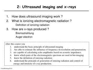
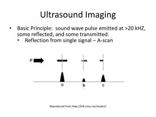
Ultrasound Imaging. Basic Principle: sound wave pulse emitted at >20 kHZ , some reflected, and some transmitted. Reflection from single signal – A-scan. Reproduced from http://folk.ntnu.no/stoylen/. B-Mode Imaging. E nergy displayed as: - amplitude, as in A-mode,

E N D
Ultrasound Imaging • Basic Principle: sound wave pulse emitted at >20 kHZ, some reflected, and some transmitted. • Reflection from single signal – A-scan Reproduced from http://folk.ntnu.no/stoylen/
B-Mode Imaging • Energy displayed as: - amplitude, as in A-mode, • - brightness, as in B-mode, • - motion curve, as in M-mode. • 2D image formed with linear or angular sweep. Reproduced from http://folk.ntnu.no/stoylen/
B-Mode 2D Imaging • Image built line by line: i) emitting the pulse, ii) waiting for reflected echoes iii) tilt beam & emit next pulse. • Cardiac application: 2D visualization of beating heart. Reproduced from http://folk.ntnu.no/stoylen/
Doppler Imaging • • Idea: moving source of sound undergoes Doppler shift. • Moving train • Blood flow: exploit frequency shift to estimate motion. Reproduced from http://folk.ntnu.no/stoylen/ & Wikimedia Commons.