Download
1 / 30
310 likes | 518 Views
Speech and Language. Lecture for the 2 nd BM course Dr Jan Schnupp jan.schnupp@dpag.ox.ac.uk. What the syllabus says you should know ( § 20.4.3-20.4.5). Core material: Mechanisms of speech production Aphasias: Wernicke’s area and sensory aphasia; Broca’s area and motor aphasia.

E N D
Speech and Language Lecture for the 2nd BM course Dr Jan Schnupp jan.schnupp@dpag.ox.ac.uk
What the syllabus says you should know (§20.4.3-20.4.5) • Core material: • Mechanisms of speech production • Aphasias: Wernicke’s area and sensory aphasia; Broca’s area and motor aphasia. • Hemispheric specialization • Extension Material: • Speech sounds and speech perception • Arcuate bundle. The dyslexias
Vocal Folds in Action http://mustelid.physiol.ox.ac.uk/drupal/?q=vocal_folds
Articulators (lips, tongue, jaw, soft palate) move to change resonance properties of the vocal tract. https://mustelid.physiol.ox.ac.uk/drupal/?q=vocalization/articulators Articulation
Harmonics and Formants of Speech Sounds Formant Harmonic
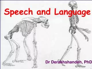
Can other animals speak? • Other mammals have similar vocal tracts and use them for communication. However, they have only very limited use of syntax (grammar) and very much smaller vocabularies than humans. • http://mustelid.physiol.ox.ac.uk/drupal/?q=mishka
Broca’s (Motor or Expressive) Aphasia • Patients with Broca’s Aphasia tend to understand speech well. • However, they have great difficulty articulating speech themselves, even though there is no severe paralysis of the articulators in the vocal tract. • Their speech tends to be halting, laboured, consisting of isolated utterances rather than full, grammatical sentences. • Suggests underlying cause is a difficulty in planning highly complicated motor acts.
A Patient with Broca’s Aphasia • From the archives of the University of Wisconsin • http://mustelid.physiol.ox.ac.uk/drupal/?q=brocas_aphasia
Broca’s Area • Broca’s aphasia is usually associated with lesion to the left frontal cortex. • See here the brain of Broca’s Patient, Mr Leborgne (“TanTan”) features a large lesion in Broca’s area.
Speech as a “Dynamic” Acoustic Signal • Most of the information in speech is carried in the manner in which the formant structure varies over time. • Pitch carries little or no semantic information (but is important in prosody and in “pitch tracking” for speech in noise. • Think of the role of the tonotopic axis in the ascending auditory pathway as representing formant distributions. (Not pitch!)
To make speech comprehensible, cochlear implants reproduce the formant structure as a pattern of electrical stimulation along the tonotopic axis of the Basilar Membrane. https://mustelid.physiol.ox.ac.uk/drupal/?q=prosthetics/noise_vocoded_speech Speech and Cochlear Implants
Spectrogram (B) and Auditory (VIII) Nerve “Neurogram” (A) of a spoken sentence. • From Delgutte B (1997) Auditory neural processing of speech. In: The Handbook of Phonetic Sciences (Laver WH, ed), pp 507-538. Oxford: Blackwell.
Auditory Cortex Neurograms of American English Onset Consonants (Engineer et al. Nat Neurosci 2008)
Where in the Brain does the Transition from Sound to Meaning happen? • We don’t really know. • “Ventral vs Dorsal stream hypothesis” of auditory cortex connectivity would suggest that anterior temporal and frontal structures should be involved. • This fits with neuroimaging studies (e.g. Scott et al (2000) Brain 123 Pt 12:2400-2406) • https://mustelid.physiol.ox.ac.uk/drupal/?q=node/46 • But other electrophysiological and lesion data do not really fit this picture.
Part 3 More about Aphasias and Clinical Observations
Receptive (Wernicke’s Aphasia) • Patients with Wernicke’s Aphasia usually have great difficulty understanding speech, even though there is no deafness. • They may speak fluently, often in long sentences, but the meaning of their sentences is unclear. (“fluent paraphasia”).
A Patient with Wernicke’s Aphasia • From the archives of the University of Wisconsin
Wernicke’s Area • Wernicke’s aphasias are often associated with lesions at the boundary of the superior temporal and parietal lobes on the left hemisphere.
Conduction Aphasia • Patients may be able to understand speech as well as produce meaningful speech, but have difficulty repeating a spoken sentence. • Often associated with damage to the Arcuate Fasciculus, which connects Wernicke’s area with frontal pre-motor structures.
The Arcuate Fasciculus Big fibre bundle connecting Broca’s and Wernicke’s Areashttp://www.biocfarm.unibo.it/aunsnc/pictef14.html
Categorizing Aphasias • Brain lesions are rarely confined to particular “classical” area boundaries. • The symptoms used to diagnose and classify aphasias can vary considerably in severity. • Thus, aphasic patients may not fit the diagnostic categories terribly well, and the way aphasias are categorized are themselves evolving.
Cortical Speech Areas and Neurosurgery • Surgeons attempting to remove epileptic foci or tumours from the brain are anxious to avoid damaging areas that are crucial for speech production or comprehension. • They may use “Wada tests” or temporary functional lesioning trough direct electrocortical stimulation. • Further reading: Calvin & Oja “Conversations with Neil’s Brain”.
Hemispheric “Dominance” for Speech and the Wada test • Broca first proposed that the left hemisphere is “dominant” for speech, based on examinations of post-mortem brains. • Nowadays “dominance” is usually assessed with the “Wada test” (intracarotid sodium amobarbital procedure): either the left or right brain hemisphere is anesthetised by injection of amobarbital into the carotid through a catheter. The patient’s ability to understand and produce speech is scored.
Left Hemisphere Dominance Dominates Wada test results suggest that: • Ca 90% of all right handed patients and ca. 75% of all left handed patients display “left hemisphere dominance” for speech. • The remaining patients are either “mixed dominant” (i.e. they need both hemispheres to process speech) or they have a “bilateral speech representation” (i.e. either hemisphere can support speech without necessarily requiring the other). • Right hemisphere dominance is comparatively rare, and seen in no more than 1-2% of the population
Hierarchical levels of speech perception • Acoustic / phonetic representation:- Can the patient tell whether two speech sounds or syllables presented in succession are the same or different? • Phonological analysis:- Can the patient tell whether two words rhyme? Or what the first phoneme (“letter”) in a given word is? • Semantic processing:- Can the patient understand “meaning”, e.g. follow spoken instructions?
“Functional Lesioning” by Electrocortical Stimulation • Sites where direct electrical stimulation can disrupt acoustic/phonetic (A), phonological (B) or semantic (C) processing of speech. • From Boatman D (2004) Cortical bases of speech perception: evidence from functional lesion studies. Cognition 92:47-65.
If you want to know more • Try chapters 1,2, 4 and 8 of “Auditory Neuroscience” by Schnupp, Nelken & King, MIT Press. • Check out auditoryneuroscience.com