Download
1 / 78
780 likes | 994 Views
NEMATODES Faculty: SAMUEL AGUAZIM, M.D. Lange Chapter 56. case. 4 year old female with constant scratching of her buttocks for 1 week. The mother says that the child has been very irritable, uncomfortable at night.

E N D
NEMATODES Faculty: SAMUEL AGUAZIM, M.D. • Lange Chapter 56
case • 4 year old female with constant scratching of her buttocks for 1 week. The mother says that the child has been very irritable, uncomfortable at night. • On examination of her anal area: numerous scratch marks, erythematous and swollen.
Pyrantel pamoate 2 doses • Recovered • Impression: Pinworm infection, secondary to Enterobius vermicularis
Nematodes • are roundworms with a cylindrical body and complete digestive tract, including a mouth and an anus. • The body is covered with a noncellular, highly resistant coating called a cuticle. • Nematodes have separate sexes; thefemale is usually larger than the male
Nematodes The medical important nematodes can be divided into two categories according to their primary location in the body to: IntestinalNematodes TissueNematodes
Intestinal Nematodes Enterobius vermicularis most common helminthic infection in the United States Disease:Pinworm infection. Characteristics:Intestinal nematode.
Enterobius vermicularis Life cycle: • confined to humans. • The infection is acquired byingesting the worm eggs. • The eggs hatch in thesmall intestine, where the larvae differentiate into adults and migrateto thecolon. • The adult male and female worms live in the colon, where mating occurs. • At night, females migrate from the anus and lay many thousands of fertilized eggs onperianal skinand in environment. • Embryo within egg becomes an infective larva within 4—6 hours. • Reinfection can occur if they are carried to themouth by fingersafter scratching the itching skin.
Enterobius vermicularis adult male and female
MOT: ingestion of eggs Females lay eggs in the perianal area at night
Enterobius vermicularis Transmission:Transmitted by ingesting eggs. Humans are the only hosts. Occurs worldwide. Pathogenesis:Worms and eggs causeperianal itching.Scratching predisposed to Secondary bacterial infection Laboratory Diagnosis:Eggs visible by “Scotch tape” technique. Eggs are not found in stool . Adult worms found in stool or near anus. Treatment:Mebendazole
Memory Tool • On the bus (Enterobius), you sit on a pin (pinworm), and get an itchy bottom and use scotch tape to make it feel better.
Trichuris trichiura Disease:Whipworminfection Characteristics:Intestinal nematode. The characteristic of“whiplike”apperance of the adult worm. Life cycle: • Humans ingesteggs,which develop into adults in gut. • Eggs are passedin feces into soil, where they embryonate, ie, become infectious.
The characteristic of “whiplike” apperance of the adult worm.
Trichuris trichiura Transmission: • More than 500 million infected. • Transmitted byfood or watercontaminated with soil containing eggs. • Humans are the only hosts. Occurs worldwide, especially in the tropics. Pathogenesis: • Worm in gut usually causeslittle damage. • The whipworm infects about 2 million children in the U.S. • Causesrectal pruritis and tenesmus, which often results inrectal prolapse.
Trichuris trichiura Laboratory Diagnosis: • Eggs visible in feces. • The egg isbarrel-shape with a plug at each end, in the stool. Treatment:Mebendazole. Prevention:Proper disposal of human waste
Trichuris trichiura eggs, a typical barrel shape two polar plugs, that are unstained
Trichuris trichiura • Tricksy (Trichuris) carries a whip and ate eggs which gave her rectal prolapse. Poor Tricksy!!!
Ascaris Disease:Ascariasis. Characteristics:Intestinal nematode. Life cycle: • Humans ingest eggs, which form larvae in gut. • Larvae migrate through the blood to thelungs, where they enter the alveoli, pass up the trachea, and areswallowed. • In the gut, they become adults and lay eggs that are passed in the feces. • They embryonate, ie, become infective in soil. • The adult worms are the largest intestinal nematodes (25 cm or more).
Ascaris Transmission:food contaminated with soil containing eggs. Humans are the only hosts.Endemic in the tropics. Pathogenesis:Larvae in lung cancause pneumonia.Heavy worm burden can cause intestinal obstruction or malnutrition. Laboratory Diagnosis:Eggs visible in feces.Eggs are oval with irregular surface. Eosinophilia occurs. Treatment:Mebendazole Prevention:Proper disposal of human waste
Billy (biliary obstruction) drives his car (Ascaris) while eatingeggs and gets short of breath. Poor Billy!!! Add intestinal obstruction
Strongyloides Disease:Strongyloidiasis Characteristics:Intestinal nematode. Life cycle: • Larvae penetrate skin, enter the blood, and migrate to thelungs. They move into alveoli and up the trachea and areswallowed. • become adults and enter the mucosa, where females produce eggs that hatch in the colon into noninfectious,rhabditiform larvaethat areusuallypassed in feces. • NOTE:the only helminth to secrete larvae (and not eggs) in feces
Strongyloides • Occasionally, rhabditiform larvae molt in the gut to forminfectious, filariform larvaethat can enter the blood and migrate to thelung (autoinfection). • The noninfectious larvae passed in feces form infectious filariform larvae in the soil. • These larvae can either penetrate the skinor form adults. • Adults in soil can undergo several entire life cycles there. • This free-living cycle can be interrupted when filariform larvae contact the skin.
Strongyloides Transmission:Filariform larvae in soil penetrate skin. Endemic in the tropics. Pathogenesis: • Little effect in immunocompetent persons. • In immunocompromised persons, massive superinfection can occur accompanied by secondary bacterial infections. Laboratory Diagnosis: Larvae visible in stool. Eosinophilia occurs. Treatment:Thiabendazole. Prevention:Proper disposal of human waste. Use of footwear
Memory Tool • The strongman (Strongyloides) is brought down by a larvae penetrating his skin causing pulmonary distress and superinfection. Poor strongman!
Ancylostoma duodenaleNecator Americanus Disease:Hookworm. Characteristics:Intestinal nematode.
Ancylostomaduodenale • Filariform larvae in soil penetrateskin of feet, enter the blood, and migrate to thelungs. • enter alveoli, pass up the trachea, then areswallowed. • become adults in small intestine and attach to walls viateeth (Ancylostoma)or cutting plates (Necator). • Eggsare passed in feces and form noninfectious rhabditiform larvae and then infectious filariform larvae.
Ancylostoma duodenale Transmission:Filariform larvae in soilpenetrate skin of feet. Humans are the only hosts. Pathogenesis:Anemia due to blood loss from gastrointestinal tract. Laboratory Diagnosis: Eggs visible in feces.Eosinophilia occurs. Treatment:Mebendazole AND Iron therapy Prevention:Use of footwear. Proper disposal of human waste.
ANCYLOSTOMA BRAZILIENSEANCYLOSTOMA CANINUM • DOG and CAT Hook worm • Forms/ Transmission. • Filariform larvae penetrate intact skin but cannot mature in humans • Disease/organ most affected • Cutaneous larvae migrans: intense itching • Tunnels through tissue • Diagnosis: clinical signs • Treatment- ivermectin
Ancylostoma braziliensis(cutaneous larva migrans/creeping eruption) Pathognomonic: serpiginous tunnels Lacks hydrolytic enzymes to penetrate into the dermis; remains localized in the epidermis
Ancylostoma braziliensis(cutaneous larva migrans/creeping eruption) Dog, cat hookworm Gulf states, South US freezing with liquid nitrogen/ethyl chloride; Mebendazole
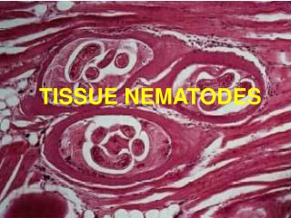
Trichinella Disease:Trichinosis. Characteristics:Intestinal nematode that encysts in tissue.
Trichinella Life cycle: • Humans ingestunder cooked pork and other meatcontaining encysted larvae, which mature into adults in small intestine. • Female worms release larvae that enter blood and migrate toskeletal muscle or brain, where theyencyst. • Pigs:most important reservoirs of human disease in USA, except Alaska where bears are the ones.Reservoir hosts are primarily pigs and rats. • Humans are dead end hosts. • Occurs worldwide but endemic in eastern Europe and west Africa
Encysted larvae of Trichinella in pressed muscle tissue. The coiled larvae can be seen inside the cysts.CDC
Trichinella Pathogenesis:Inflammation ofmuscle Laboratory Diagnosis: • Encysted larvae visible in muscle biopsy • Eosinophilia • Serologic tests positive. Treatment:Thiabendazole effective early against adults. None for established disease Prevention:Adequate cooking of pork
Memory Tool • A tricky (Trichinella) pig caused cysts in his owners muscles.
Tissue nematodeDracunculus Disease:Dracunculiasis. Characteristics:Tissue nematode.