紫草素促傷口癒合之離體細胞培養以及活體動物試驗: SMAD 蛋白訊息傳遞路徑在紫草素誘發前膠原蛋白生成中所扮演之角色
紫草素促傷口癒合之離體細胞培養以及活體動物試驗: SMAD 蛋白訊息傳遞路徑在紫草素誘發前膠原蛋白生成中所扮演之角色.

紫草素促傷口癒合之離體細胞培養以及活體動物試驗: SMAD 蛋白訊息傳遞路徑在紫草素誘發前膠原蛋白生成中所扮演之角色
E N D
Presentation Transcript
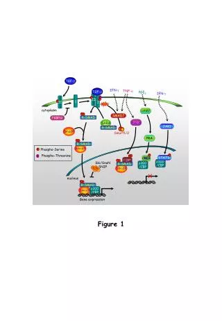
紫草素促傷口癒合之離體細胞培養以及活體動物試驗:SMAD 蛋白訊息傳遞路徑在紫草素誘發前膠原蛋白生成中所扮演之角色 • 皮膚是人體最大的器官,由多層上皮組織構成,覆蓋於整個人體,形成屏障保護人體免於外來致病原。是故皮膚對人體而言相當重要。當人體受傷時,會引發一連串的表皮創傷癒合過程,以修復並治癒傷口,進行真皮與上皮組織的再建構,為人體的自然修復過程。通常此過程可分為三階段互相重疊的時期,包括發炎反應、組織增生和組織重建期。紫草素是由紫草(Lithospermum euchroma, Boraginaceae)之根萃取出的主要成份。在古代中國與日本入藥為「紫雲膏」來治療刀傷與燒燙傷。許多文獻指出,紫草素具有許多生理活性,包括抗發炎、抗菌、細胞凋亡、抗血管新生、抗腫瘤及促肉芽組織增生等。肉芽組織生成與膠原蛋白堆積,為創傷癒合過程的組織增生期中,至為重要的關鍵步驟。而人類纖維母細胞為肉芽組織和生成膠原蛋白的主要細胞。在本文裡,我們分別透過活體動物實驗及離體試驗,探討紫草素在促進創傷癒合過程中,對於人類皮膚纖維母細胞的影響。在活體動物實驗中,我們利用皮膚切片法,在小鼠背部創造出直徑6 mm 的傷口。每天局部給予紫草素、bFGF(basic Fibroblast growthfactor)當正向控制組,PEG(Polyethylene glycol)為對照組。給藥期間為14 天,每天觀察測量傷口並記錄面積。由實驗結果發現,給予bFGF正向控制劑的組別擁有最顯著的癒合效果,另外,給予0.1%紫草素的組別相較於對照組,促進傷口癒合的能力亦明顯較高,代表紫草素參與了傷口癒合的過程。為了進一步研究紫草素促進創傷癒合的機轉,我們選用人類纖維母細胞細胞株(CCD-966SK)作為實驗細胞。在離體細胞培養實驗裡,紫草素能夠誘發人類纖維母細胞的細胞遷移現象及ERK1/2 蛋白質(Extracellular signal-regulated kinase)磷酸化,且呈現濃度相關。ERK磷酸化通常與細胞增生反應有關。此外,紫草素亦能誘發MMP-1(Matrix Metalloprotease-1)的蛋白質表現及抑制PAI-1(Plasminogenactivator inhibitor type I)蛋白質表現,也呈現濃度相關性。MMP-1 與PAI-1 同時在ECM(Extracellular matrix)降解、纖維蛋白溶解和細胞遷移中扮演重要的角色。另一方面,近年來SMAD3 路徑被認為是調控創傷癒合的路徑之一,在紫草素的作用下,TGF-beta /SMAD 路徑中的分子,SMAD3(Small mother against decapentaplegic)和procollagen也會在人類皮膚纖維母細胞中被誘發出來。綜合以上所述,從實驗結果推測,紫草素透過ERK 的磷酸化、procollagen、PAI-1 和SMAD 的蛋白質表現,能加速創傷癒合,而MMP-1 亦參與其中。MMP-1 在創傷癒合過程中的定位仍有所爭議,但也有報導指出,MMP-1 的誘發與創傷癒合有關。因此我們認為,紫草素在活體動物實驗與離體細胞試驗中,會透過SMAD 訊息傳遞路徑,促使傷口癒合。而這些結果也提供了我們更多資訊,助於發展藥理臨床上創傷治療的應用。
Effect of shikonin on promoting wound healing in vitro andin vivo : the role of SMAD signaling in shikonin induced procollagen formation • Skin is the largest organ of our bodies, and made up of multiplelayers of epithelial tissues that cover the bodies protect us from theexternal pathogens, also considered is one of the most important parts inhuman organs. While an individual injuries, a set of events calledcutaneous wound healing will take place to repair the damage. It is anatural process of regenerating dermal and epidermal tissue, andcategorized into several overlapping steps: the inflammatory, proliferative,and maturation phases.Shikonin is a major components isolated from the root ofLithospermum euchroma (Boraginaceae). It was used as an ointmentcalled SHIUNKO for treating carbuncles and burns in China and Japanfor a long time. Many studies showed that shikonin demonstrated manybiological effects such as anti-inflammatory, antimicrobial, apoptotic,anti-angiogenesis, anti-tumor, and proliferation of granulation tissue.Since the granulation tissue formation and collagen deposition arecritical events of the proliferation phase of wound healing. In this study,we investigated the effects of shikonin on human skin fibroblast onpromoting wound healing in vitro and in vivo.In in vivo study, mice were created a 6 mm diameter wound on backby biopsy punch. Topical treating with shikonin、bFGF (basic Fibroblastgrowth factor)(positive control) and PEG(Polyethylene glycol)(negativecontrol) for 14 days, then measured and observed the wound area andrecorded everyday. Our data suggested bFGF treated groups showedlargely healing promoted effect and taken as positive control, 0.1%shikonin also significantly promoted wound healing compared withcontrol mice. This data indicated the involvement of shikonin in woundhealing.In order to investigate the mechanism of shikonin in promoting thewound healing, we use human skin fibroblast cell culture line(CCD-966SK) in our study. In in vitro cell culture experiments, we foundshikonin could concentration-dependently induce cell migration andERK1/2(Extracellular signal-regulated kinase) phosphorylation in humanskin fibroblasts. ERK phosphorylation is general accepted by contributingto cell proliferation. In addition, shikonin also concentratio-dependentlyincreased MMP-1(Matrix metalloproteinase-1) induction, and decreasePAI-1(Plasminogen activator inhibitor type I) inhibition. Both MMP-1and PAI-1 play an important role for ECM (Extracellular matrix)degradation, fibrinolysis and cell migration. On the other hand, themolecule in TGF-beta/SMAD pathway, SMAD3 (Small mother againstdecapentaplegic), and procollagen were all increased in shikonin treatedskin fibroblast cells. SMAD3 pathway is recognized in controlling thehealing pathway in recent years.Take together, our data indicated, shikonin could accelerate woundhealing by promoting ERK phosphorylation and procollagen、PAI-I and SMAD protein induction, MMP-1 also involve in this effect. Although the role of MMP-1 in wound healing is controversial, but still have reports suggested the increase of MMP-1 is involved in the pathway. We concluded, shikonin can promote wound healing both in vitro and in vivo through SMAD signaling pathway, and this finding can provide the information for pharmacological use in the clinical care of wound healing.