Download
1 / 27
370 likes | 942 Views
Functional Organization of the Cardiovascular System. Objectives. Describe the functional organization of cardiovascular system List the functions of cardiovascular system. Describe the main function of arteries, capillaries and veins

E N D
Objectives • Describe the functional organization of cardiovascular system • List the functions of cardiovascular system. • Describe the main function of arteries, capillaries and veins • Describe the flow of blood through the chambers of the heart and through the systemic and pulmonary circulations. • Compare and contrast the systemic and pulmonary circulation.
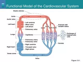
HEART (PUMP) CARDIOVASCULAR SYSTEM VESSELS (DISTRIBUTION SYSTEM) Blood Functional Organization of Cardiovascular system
Functions of Cardiovascular System: I. Primary (main) function of the heart: ♥ Acts as a muscular pump: in order to maintain adequate level of blood flow throughout CVS by pumping blood under press into vascular system. ♥ Responsible for the mass movement of fluid in body.
Functions of Cardiovascular System (continued) II. Secondary functions: 1. Transportation: delivers O2 to tissues, & brings back CO2 to lungs. carries absorbed digestion products to liver & tissues. carries metabolic wastes to kidneys to be excreted. distribution of body fluids. 2. Regulation: Hormonal: carries hormones to target tissues to produce their effects. Immune: carries antibodies, leukocytes (WBCs), cytokines, & complement to aid body defense mechanism against pathogens. Protection: carries platelets, & clotting factors to aid protection of the body in blood clotting mechanism. Temperature: helps in regulation of body temperature, by diverting blood to cool or warm the body.
Anatomy of the heart: • Positioned between two bony structures – sternum and vertebrae (CPR) • Hollow, muscular organ.
Vena cava pulmonary veins Pulmonary trunk (Aorta)
Atrium: weak primer pump for the ventricle Ventricle: the main pumping force Rt. Ventricle Lt. ventricle Pulmonary circulation Systemic circulation
systemic circulation Pulmonary circulation
Valves of the heart: ♥2 atrioventricular (AV) valves: ■One way valves. ■Allow blood to flow from atria into ventricles. ■ Tricuspid (Rt) & Mitral (Lt). ♥2 semilunar valves: ■One way valves. ■At origin of pulmonary artery & aorta. ■ Pulmonary (Rt) & Aortic (Lt). ■ Open during ventricular contraction.
Heart Valves • One way flow in heart is ensured by heart valves • Valves open & close passively - open by forward P by blood - close by backward P by blood
No valves between atria and veins • Reasons • Atrial pressures usually are not much higher than venous pressures • Sites where venae cavae enter atria are partially compressed during atrial contraction
Serves 3 roles: A mechanical base: atria anchored above and ventricles below Perforated by 4 apertures, each containing a valve Insulates the ventricles The fibrous skeleton of the heart
Vascular Tree • Closed system of vessels • Consists of • Arteries • Carry blood away from heart to tissues • Arterioles • Smaller branches of arteries • Capillaries • Smaller branches of arterioles • Smallest of vessels across which all exchanges are made with surrounding cells • Venules • Formed when capillaries rejoin • Return blood to heart • Veins • Formed when venules merge • Return blood to heart
Arteries • Function: • Rapid transit passage-ways for blood from heart to tissues • Pressure reservoir Structure of arterial wall • Plentiful of elastic fibers….high compliance
Arterioles (resistance vessels) • Very small arteries that delivers blood to capillaries • Structure • Very little elastic tissue but thick layer of smooth muscle • Function Regulating blood flow from arteries to capillaries by regulating resistance according to tissue metabolic needs.
Capillaries • Microscopic vessels that connects arterioles to venules • Structure • Single wall layered vessels (endothelial cells) • Undergoes extensive branching • Maximized surface area and minimized diffusion distance • Velocity of blood flow through capillaries is relatively slow • Provides adequate exchange time • Function: • Exchange of nutrients and wastes between blood and tissue cells
Capillaries cont. • Under resting conditions many capillaries are not open • Capillaries surrounded by precapillary sphincters • Contraction of sphincters reduces blood flowing into capillaries in an organ • Relaxation of sphincters has opposite effect
Veins • Carry blood from tissues to heart • Structure: • Thin wall • Less smooth muscle and considerable amount of collagen • Less elastic fibers • Function: • Passage ways back to heart • Blood reservoir (capacitance vessels)