Download
1 / 15
150 likes | 306 Views
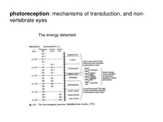
photoreception : mechanisms of transduction, and non-vertebrate eyes. The energy detected:. The game plan. Who does it… or where do you find sensory organs containing retinal / rhodopsin?.

E N D
photoreception: mechanisms of transduction, and non-vertebrate eyes The energy detected:
Who does it… or where do you find sensory organs containing retinal / rhodopsin? Note: there is also Bacteriorhodopsin – similar to rhodopsin of all other organisms - but not used for photo-sensory detection. Rather it is a mechanism like photosynthesis to drive uni-directional ion transport for energy generation.
Retinal in opsin pocket (four different 11-cis isomers) From: Jang et al, Mechanism of rhodopsin activation as examined with ring contstrained retinal analogs and the crystal structure of the ground state protein. J. Biol. Chem. (2001) 276:26148-26153
Structural shift due to photo absorption Rhodopsin, shown in the resting state (magenta, top view), is the photoreceptor protein of the vertebrate retina. Photoisomerization of the bound retinal chromophore (red) triggers a rigid-body helix tilt of a single helix (yellow), as dramatically demonstrated by a 6 Å increase in the distance between nitroxide spin labels attached to sites 63 and 241 (stick models). The distances were measured using DEER (Jeschke, Chemphyschem 2002, 3, 927) on an ELEXSYS E580 spectrometer. http://www.bruker-biospin.com/brukerepr/october.html
The photoisomerization of retinal (“primary process”, next page) is believed to be complete in only 100---200 fs, making it one of the fastest known photoreactions [Q. Wang, R.W. Shoenlein, L.A. Peteanu, R.A. Mathies, and C.V. Shank, Science 266, 422 (1994)].
In vertebrate photoreceptor cells, activated retinal begins a biochemical cascade that changes transmembrane ion permeability.
In vertebrate photoreceptor cells, activated retinal begins a biochemical cascade that changes transmembrane ion permeability. 5’GMP cGMP Rh Rh* Keeps ion channel open Reduce cGMP pool opsin Δ conformation PDE* opsin’s binding site for G-protein (transducin) exposed The mobile Tα-GTP binds to phosphodiesterase (some may also diffuse through cytosol) Bound transducin’s α-Subunit can no longer hold on to GDP, so it falls off. Once GTP replaces GDP, the subunit Tα-GTP separates from the rest of transducin α-Subunit conformation now can bind GTP in place of GDP
The proteins activated interact in the endomembrane (mostly)


![[VII]. Regulation of Gene Expression Via Signal Transduction](https://cdn2.slideserve.com/4429639/slide1-dt.jpg)