Download
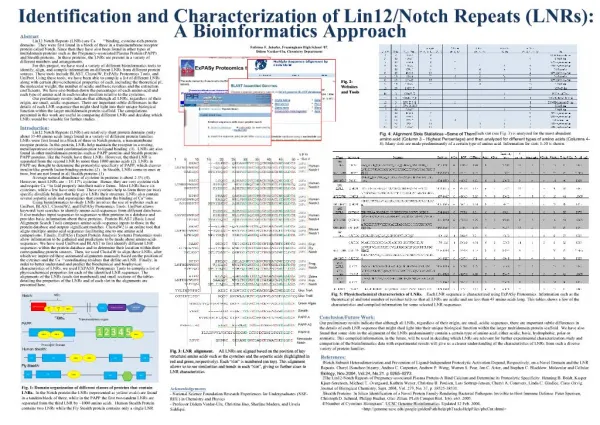
1 / 28
310 likes | 593 Views
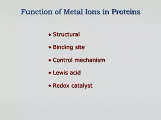
Identification and Characterization of Metal Ions in Proteins. Determine the presence of metal ions Determine a role for metal ions remove the metal ion replace the metal ion identify the binding environment identify the function. How can we accomplish this ?.

E N D
Identification and Characterization of Metal Ions in Proteins • Determine the presence of metal ions • Determine a role for metal ions • remove the metal ion • replace the metal ion • identify the binding environment • identify the function How can we accomplish this ?
Physical Methods to Characterize Metal Complexes • Presence of metal ions in biomolecules • Atomic absorption spectroscopy • UV-visible spectroscopy • Fluorescence spectroscopy • Environment of metal ions in biomolecules • Low resolution methods • UV-visible spectroscopy • Fluorescence spectroscopy • CD spectroscopy • Mossbauer spectroscopy • Medium resolution methods • EPR spectroscopy • EXAFS • High resolution methods • NMR spectroscopy • X-ray diffraction
Time scale of spectroscopic techniques very fast fast • time scales are determined by the frequency of light • very fast reactions (e.g. electron transfer reactions) require very fast spectroscopic methods to follow the reactions
Atomic Absorption Spectroscopydetectable elements All of the biologically relevant elements can be detected by atomic absorption spectroscopy
Atomic Absorption Spectroscopyinstrument block diagram each element requires its own unique lamp graphite furnace atomization flame atomization
UV-visible spectroscopy • The presence of Fe, Co, Ni and Cu can be detected by visible spectroscopy • In favorable cases some geometric information can be obtained
UV-visible spectroscopyhuman carbonic anhydrase The metal ion environment is sensitive to inhibitor binding
UV-visible spectroscopyCo(II) complexes Tetrahedral Co(II) complexes have higher transition probabilities (greater molar absorbance) with lower transition energies (higher wavelengths)
Fluorescence spectroscopy • excitation to a relatively long lived excited state allows sufficient time for vibrational relaxations • when electrons decay back to the ground state the transition occurs at lower energy (higher wavelength)
Circular Dichroism Circularly polarized light contains both left-handed and right-handed components Chiral molecules (such as amino acids) will absorb one component more strongly than the other As a consequence there will be a rotation in the plane of polarized light
Circular Dichroism For proteins both the direction and the magnitude of the rotation will be influenced by the secondary structural elements
Mossbauer spectroscopy Most useful to examine the oxidation states of iron in biological complexes The parameters in a Mossbauer spectrum are the isomer shift (δ) and the splitting (∆Eo)
EPR spectroscopy Measures the environment of unpaired electrons in paramagnetic complexes The parameters in an EPR spectrum are the peak positions (g values) and the hyperfine splitting (A values) Distortions in the spectra contains information about the coordination geometry
EPR spectroscopy • The EPR spectra of Mn(II) complexes are quite different, but can also contains information about: • coordination geometry • donor atom types
EPR spectroscopy Many biologically important metal ions produce useful EPR spectra
EXAFS Extended X-ray absorption fine structure is a scattering technique that can detect heavy atoms in the vicinity of a metal center • In the best cases EXAFS scattering can give information about: • the types of donor atoms • the number of donor atoms • the metal-donor atom distances
EXAFS single shells multiple shells
NMR spectroscopy Two-dimensional spectra are used to assign resonances to different atoms in the structural and to determine their position relative to the other atoms
NMR spectroscopy A more useful presentation is to look at the spectrum from a top view The off diagonal peaks (cross peaks) show the close through-space contacts between different atoms
NMR spectroscopy An expansion of the NH spectral region shows the assignments for each individual amino acid in the protein Once each peak has been assigned then the presence of cross peaks will indicate the nearest neighbors for each hydrogen atom
NMR spectroscopy A map of all of the nearest neighbor distances will produce a three-dimensional structure of a protein Different pulse sequences will give different information about the environment of each nuclear spin
X-ray Diffractionbasic principles An intense beam of x-rays is focused on a protein crystal A small fraction of these x-rays are diffracted by interactions with the electrons that surround each atom in a protein molecule The location and intensity of the spots in the diffraction patterns contain information about the location of the atoms in the protein molecule
X-ray Diffraction diffraction pattern Both the position and the intensity of each spot contains information about the location of the atoms in the molecule The greater the angle of diffraction the more precisely determined are the atom positions
Metal ion probes Not all biologically important metal ions have spectral signals that can be detected by the methods discussed
Summary • Atomic absorption, UV-visible and fluorescence spectroscopy can be used to detect the presence of metal ions in biomolecules • UV-visible, fluorescence, CD and Mossbauer spectroscopy can provide some information about the environment of the bound metal ion • EPR and EXAFS can identify the nature of the donor atoms and the metal ion coordination geometry • NMR and X-ray diffraction studies will produce a high-resolution structural picture of biological macromolecules