Download

1 / 6
60 likes | 73 Views
Explore the causes and effects of magnetic field inhomogeneity artifacts in brain imaging, with a focus on ASL and DTI images. Learn how susceptibility and coil issues can impact image quality.

E N D
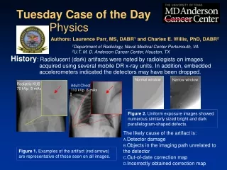
History: Tuesday Case of the Day • The likely cause of the artifact is: • Malfunctioning receive coil • Transmit (B1) inhomogeneity • Susceptibility-related T2* dephasing • Magnetic field (B0) inhomogeneity Physics Authors: Dustin K. Ragan, PhD1 and Charles E. Willis, PhD, DABR2 1Department of Pediatrics, Washington University, St. Louis, MO 2U.T. M. D. Anderson Cancer Center, Houston, TX Substantial signal dropout was observed in arterial spin label (ASL) images, acquired at 3T in a young female patient three-months after mild traumatic brain injury Figure 2. No artifacts are displayed on the DTI images Figure 1. Examples of the artifact (red arrows) which was only observed in this one sequence and this one patient
1. The same coil was used to acquire all images, but signal loss is only visible in one sequence 2. DTI images are also typically acquired with an EPI-readout, which would also be sensitivity signal dropout from magnetic susceptibility in the brain 3. Artifact is located at the periphery of the brain Findings: Anatomical and DTI images did not display signal loss Figure 4: DTI image without artifact Figure 5: T1-weighted anatomical image Figure 3: ASL image with artifact
Coil problems would manifest across all of the acquired images. Therefore, choice A. Malfunctioning receive coil is incorrect. Transmit inhomogeneity typically produces shading on the interior of the images, not at the periphery. Therefore, choice B. Transmit (B1) inhomogeneity is incorrect. The patient was wearing a shirt with iron-based glitter up to the shoulder. This distorted the position of the labeling pulses, causing them to saturate brain tissue instead of blood. (Magnetic field inhomogeneties in the brain can distort both ASL and DTI, as in the figure, but only ASL is sensitive to the neck) Susceptibility-related dephasing is common in EPI acquisitions, which is used in both ASL and DTI acquisitions. Because only the ASL images were affected, choice C. Susceptibility-related dephasing is incorrect. Also, large susceptibility artifacts are relatively rare in mild TBI, so it is inconsistent with the patient’s presentation. Discussion: Pseudocontinous ASL uses a preparatory train of RF pulses positioned at the neck along with (usually) an EPI readout to acquire images of cerebral blood flow Figure 6: Susceptibility artifacts present on both ASL and DTI images
Discussion: The gradient strengths used in ASL labeling are relatively weak, which amplifies the distorting effect of field inhomogeneities ΔB=Gx A magnetic field shift produces a much larger distortion in the presence of a weak gradient than a strong one Gradient strengths used slice selection are around ~40 mT/m; those used in labeling are ~10 mT/m As a result, the ASL labeling pulses are highly susceptible to position shifts RF pulses are not localized and can unintentionally affect the entire sensitive volume of the coil Strong gradient Magnetic field offset Weak gradient Physical position
References/Bibliography: Jahanian H, et al. B0 field inhomogeneity considerations in pseudo-continuous arterial spin labelng (pCASL): effects on tagging efficiency and correction strategy. NMR in Biomedicine 24: 1202-1209. 2011. Haacke M., et al. Magnetic Field Inhomogeneity Effects and T2* Dephasing, In Magnetic Resonance Imaging: Physical Principles and Sequence Design. p. 569-617. New York, John Wiley & Sons.