Download
1 / 57
570 likes | 691 Views
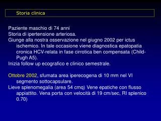
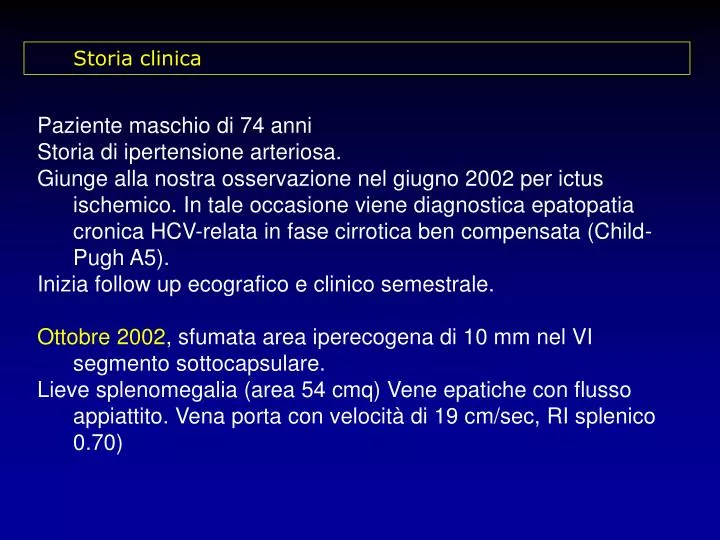
Storia clinica. Paziente maschio di 74 anni Storia di ipertensione arteriosa. Giunge alla nostra osservazione nel giugno 2002 per ictus ischemico. In tale occasione viene diagnostica epatopatia cronica HCV-relata in fase cirrotica ben compensata (Child-Pugh A5).

E N D
Storia clinica Paziente maschio di 74 anni Storia di ipertensione arteriosa. Giunge alla nostra osservazione nel giugno 2002 per ictus ischemico. In tale occasione viene diagnostica epatopatia cronica HCV-relata in fase cirrotica ben compensata (Child-Pugh A5). Inizia follow up ecografico e clinico semestrale. Ottobre 2002, sfumata area iperecogena di 10 mm nel VI segmento sottocapsulare. Lieve splenomegalia (area 54 cmq) Vene epatiche con flusso appiattito. Vena porta con velocità di 19 cm/sec, RI splenico 0.70)
Viene rivisto a 4 mesi circa (febbraio 2003). Si conferma il piccolo nodulo di 11 mm. Si consiglia TC. Viene eseguita e risulta negativa. Si programma uno stretto follow up. • Maggio 2003Permane immodificata la lesione debolmente iperecogena di 11 mm nel VI segmento. Non ulteriori lesioni focali. • Ottobre 2003In sede centroepatica, strettamente adiacente al ramo portale posteriore destro, è presente un'area ipoecogena di 17 mm con scarsi segnali vascolari al suo interno.Permane invariata la lesione focale debolmente iperecogena di 11 mm al 6° segmento. Si procede ad angioecografia perfusionale.
TC spirale trifasica (26.11.2003) Fase arteriosa Fase portale
TC spirale trifasica (26.11.2003) ? Fase portale
CLINICA, DIAGNOSTICA E TERAPIA DELL’EPATOCARCINOMA Luigi Bolondi Cattedra di Clinica Medica Dipartimento di Medicina Interna e Gastroenterologia Università di Bologna - Policlinico S. Orsola Malpighi “
INCIDENCE OF HCC IN LIVER CIRRHOSIS annual incidence Oka et al, 1990 6.5 % Colombo et al, 1991 3 % Pateron et al, 1994 5.8 % Benvegnu et al, 1994 3 % Cottone et al, 1994 1.5-10 % Solmi et al, 1996 1.4 % Bolondi et al, 2001 4.1 %
CIRROSI VIRUS HCC Diminuita capacità riparatrice dei danni al DNA Flogosi cronica Necrosi Rigenerazione epatocitaria HCC Aumento errori di replicazione e trascrizione del DNA Eterogeneità geografica Diversi fattori di rischio Diversi bersagli a livello molecolare
Factors affecting natural history Acute hepatitis Chronic hepatitis HLA type 85% Male gender Age on onset Alcohol Interferon Cirrhosis 20% Hepatitis B Alcohol Interferon 6% Decompensation HCC 4% Transplantation Death 3,6% Di Bisceglie, Hepatology, 2000
INCIDENCE OF HCC DURING THE SURVEILLANCE PROGRAMME OF LIVER CIRRHOSIS (1989-1997) 313 patients with a follow-up of 56 31 months 74 nodules (23,6 %) 13 cases non HCC 61 HCC (19,5 %) Bolondi et al. Gut 2001
SCREENING FOR HCC IN CIRRHOSIS ANALYSIS OF SURVIVAL BENEFIT Significant longer survivals for screened vs non screened p = 0.009 (Wong, Liver Transpl 2000) p < 0.0001 (Yuen, Hepatology 2000) p < 0.02 (Bolondi, Gut 2001) p < 0.001 (Trevisani, Am J Gastro 2002) No Significant difference * (Sarasin, Am J Med 1996) * transplantation not included in the model
Tailoring screening on RISK FACTORS FOR HCC IN CIRRHOSIS • Age(Aizawa, Cancer 2000) • Male gender(Zoli, Cancer 1996 • Bolondi, Gut 2001 • El Serag, J Clin Gastro 2002) • Child-Pugh score(Bolondi, GUT 2001) • HBsAg +(Solmi, Am J Gastro 1996) • Tsukuma, N Engl J Med 1993) • HCV+(Velazquez, Hepatology 2003) • HBV + HCV(Parkin, IARC 1992) • HCV + alcol(Benvegnù, Gut 2001) • AFP(Bolondi, Gut 2001)
DEVELOPEMENT OF NEOPLASTIC GROWTH IN MACROREGENERATIVE NODULES ARAKAWA 1986 RECOGNITION OF EARLY MALIGNANT FOCI IN 5 ADENOMATOUS HYPERPLASTIC NODULES N°nodules mean follow-up 9 neoplastic growth TAKAYAMA 1990 18 1-5 yrs 9 benign behaviour 10 neoplastic growth RAPACCINI 1990 12 10.2 mos 2 benign behaviour 0 neoplastic growth KONDO 1990 17 > 1 yr 17 benign behaviour 7 neoplastic growth BOLONDI 1993 12 22.6 mos 5 benign behaviour
PREDICTION OF MALIGNANT EVOLUTION IN SMALL NODULES (< 1.5 cm) DETECTED AT IMAGING TECHNIQUES IMAGING NEW TISSUE MARKERS MOLECULAR ANALYSIS • Markers of proliferation (AgNORs, PCNA, Ki67...) • Enzymatic cytochemistry • DNA ploidy • Assessment of monoclonality • Genomic instability and LOH • Assessment of vascularity Probably no consequence on outcome CLINICAL CRITERIA Volume increase at 4 month
Blood supply of liver nodules in cirrhosis Portal flow Arterial flow Large regenerative nodule HCC Dysplastic nodule Borderline lesion
DOPPLER QUANTITATIVE QUALITATIVE SPIRAL CT SPECTRAL ANALYSIS COLOR and POWER mapping + mdc Contrast-enhanced NMR CONTRAST-ENHANCED US Stimulated Acoustic Emission Harmonic Imaging Pulse Inversion C3-mode CnTi CHARACTERIZATION OF LIVER MASSES: ASSESSMENT OF VASCULARITY BY IMAGING TECHNIQUES
ARTERIAL HYPERVASCULARITY IN SMALL HEPATOCELLULAR CARCINOMA Perfusional Angiosonography with Sonovue Spiral CT enhanced artherial phase
HCC - Hyperintensity in the arterial phase - Iso or Hypointensity in the portal and late phases
DIAGNOSIS OF HCC Cirrhotic patients (US + AFP/6m) Liver nodule No nodule 1-2 cm > 2 cm < 1 cm Increased AFP* Normal AFP FNAB US /3m Spiral CT AFP > 400 ng/ml Doppler/CT/MRI/An HCC No HCC Surveillance US + AFP/6m * AFP level >200ng/dl Bruix, J Hepatol ,2001
STAGING:OPEN PROBLEMS AND AREAS FOR FUTURE RESEARCHES • Multinodularity • Vascular invasion Selection between radical treatment or palliation Imaging techniques insufficient • Selection between OLT and ablation/destruction therapies • Need for adjuvant therapy • Recurrence potential Tissue and molecular markers (Currently not done)
THERAPEUTIC OPTIONS FOR HCC Local therapy Surgical resection Transplant Percutaneous echo-guided Intra-arterial Systemic chemotherapy or hormonal therapy
EFFECT OF TREATMENT ON SURVIVAL OF 1108 PTS WITH HCC Multicentric Italian Study Group on HCC SURVIVAL OF SINGLE HCC <5 cm Child A J Hepatol, 1995
SCREENING FOR HCC IN CIRRHOSIS ELIGIBILITY FOR CURATIVE TREATMENTS HCC detected outside surveillance programme HCC detected within surveillance programme 47.5 % 31.7 % p < 0.01 (Bolondi, Gut 2001)
Rationale for the use of local treatments • High rate of exclusion criteria from surgical resection • (5-9% of pts arising from screening are • candidate to surgery) • High recurrence rate after surgical resection • 3 year recurrence 72% Ikeda et al, 1993 • 5 year recurrence 83% Ng et al, 1995 • 100% Belghiti et al, 1991 • 91% Gouillat et al, 1999
HEATlaser, radiofrequency, highly focused ultrasound • FROST cryosurgery • DRUGS alcohol injection • RADIOACTIVITY implantation of radioactive seeds INTERSTITIAL TUMOR ABLATION
Survival (%) No. of Pts Author and year 1-yr 3-yr 5-yr Shiina S et al, AJR 1993 Livraghi T et al, Radiology 1995 Child A, single < 5 cm Child B, single < 5 cm Lencioni R et al, Cancer 1995 Child A, single / multiple < 3 cm Child B, single / multiple < 3 cm 98 293 149 64 41 85 98 93 100 91 62 79 63 87 53 52 47 29 55 13 Survival Outcomes in PEI-Treated Pts (Retrospective Studies)
Surgery > PEI (n=8.010) (n=4.037) SURVIVAL AFTER SURGICAL AND NONSURGICAL TREATMENT FOR HCC HCC < 2 cm clinical stage I 5 cm > HCC > 2 cm all clinical stages (retrospective study) (Arii et al, Hepatology 2000 Liver Cancer Study Group of Japan)
PEI versus Surgical Resection (Non-Randomized Studies) p = N.S. p = N.S. - Same tumor stage - Poorer liver function in PEI groups Yamamoto et al, Hepatology 2001 Castells et al, Hepatology 1993
Okosa farmaka ouk ihtai, sidheros ihtai, osa sidheros ouk ihtai, pur ihtai, osa dh pur ouk ihtai tauta crh nomizein aniata Quae maedicamenta non sanant, ferrum sanat,quae ferrum non sanat, ignis sanat,quae vero ignis non sanat, insanabilia reputari oportet Ippocrate, Aforisma 7, 87
RF THERMAL ABLATION EXPANDABLE NEEDLE (1.9 mm) 90-115°C 4 to 10 nickel-titanium hooks with tip thermistors
RF THERMAL ABLATION COOLED-TIP NEEDLE (1.2-1.3 mm) 20-25°C Peristaltic pump with 0°C saline solution
24 pts, 47 HCC lesions (0.4 – 5.5 cm; mean, 2.3 cm) - Complete necrosis on histology: 35 / 47 (74%) RF Ablation of HCC: Local Effect (histologic assessment after OLT) Lu DSK et al, Radiology 2005
Overall Survival 67% PEI series (n = 184) - Lencioni R et al, Eur Radiol 1997
74% 50% Lin SM et al Gastroenterology 2004
RANDOMIZED COMPARISON OF RF THERMAL ABLATION vs PEI232 patients with up to 3 HCC < 3 cm each RF PEI ---------------------------------------------------------------- • Treatment sessions 2.1 6.4 p<00001 • 4yr survival 74% 57% p=0.01 • 4yr Overall recurrence 70% 85% p=0.005 • 4yr Local progression 1.7% 11% p=0.003 Shiina, Gastroenterology 2005
COMPARING THE OUTCOMES OF RF ABLATION AND SURGERY IN PTS WITH SINGLE SMALL HCC AND WELL-PRESERVED HEPATIC FUNCTION Hong SN et al, J Clin Gastroenterol 2005
Barcelona 2005 - PERCUTANEOUS ABLATIONSummary and conclusions • RF thermal ablation has emerged as the most valid alternative to PEI. According to various studies, its failure in achieving local control is lower than PEI. Data on survival are still preliminary and are influenced by different patient selection • The complication rate of RF was initially considered higher but recent reports do not confirm this finding • In HCCs of 3 to 5 cm the efficacy of a percutaneous treatment in achieving local control is questionable • Individual factors play an important role in treatment selection • Other techniques such as microwave or Laser have a minor impact • PEI can probably maintain a place in the treatment of very small nodules (<2 cm) or in difficult locations (perivascular)
PROBLEMS IN EVALUATION RCTs ON TRANSARTERIAL CHEMOEMBOLIZATION • Small sample size • Differences in treatment procedures (chemoterapeutic agent - Cysplatin, Mytomicin, Doxorubicin - , embolization, number and interval of procedures) • Patients selection and stratification
TERAPIA DELL’ HCC UNIFOCALE IN FEGATO CIRROTICO CONCETTI CHIAVE Pz in classe Child-Pugh A a basso rischio operatorio e nodulo unico candidati a resezione anatomica Pz con nodulo singolo < 5 cm (e buon compenso epatico) ottimi candidati alle terapie locoregionali percutanee: l’alcolizzazione è la tecnica di scelta Noduli < 3 cm: Risultati migliori Noduli >3 cm: Se non resecabili, si può associare PEI + TACE
TERAPIA DELL’HCC UNIFOCALE IN FEGATO CIRROTICO CONCETTI CHIAVE Pz < 65 aa con nodulo singolo in classe Child-Pugh B e C Considerare indicazione a trapianto di fegato La TACE può essere utile nei pz in lista d’attesa per contrastare la crescita e la diffusione della neoplasia (?)
BARCELONA RECOMMENDATIONS CURATIVE TREATMENTS PEI vs SURGICAL RESECTION Recurrence rate after percutaneous treatments is as frequent as after surgical resection (>50% at 3 years and > 70% at 5 years) The are no RCTs comparing surgical resection and PEI. While some series report that survival after PEI is lower than after surgical resection, some cohort studies have failed to detect significant differences PEI can be recommended for well compensated patients when surgery is precluded J Hepatol 2001