Download
1 / 43
680 likes | 1.52k Views
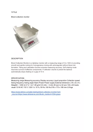
Blood Collection and Handling of Blood Samples. Collecting your Sample. Determine which ________________ are needed. Determine the __________________ you will need and the _______________ you will use.

E N D
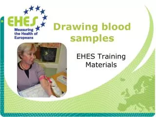
Collecting your Sample • Determine which ________________ are needed. • Determine the __________________ you will need and the _______________ you will use. • Preferred blood source is almost ALWAYS _______________ blood, not ______________ blood. ___________ vein is usually most appropriate vessel for collection. • Use the _________________needle that the patient can comfortably accommodate. • Choose the size syringe that best matches the ___________________________________ you will need.
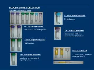
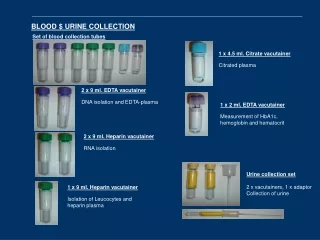
The Vacutainer • Is composed of a ______________, _________________________and _________________tubes. • Use the correct size tube to minimize _______________of the sample and to prevent ____________________ of the vein. • Fill tube to correct volume based on strength of ______________________________ to ensure appropriate ratio of blood to anticoagulant. • ADVANTAGE: multiple samples can be collected directly into tubes without __________________________________ from patient.
Sample Volume • The amount of blood collected from an animal depends on the amount of _______________or _______________needed as well as the ________________________ of the animal. • Enough blood should be taken to run the required tests _______ times. This should be enough to compensate for technician error, instrument error or the need for diluted samples.
Serum or Plasma? • Serum or plasma are the _____________ portion of whole blood. • Fluid portion of blood is ____% water, ____% dissolved constituents like proteins, vitamins, carbs, hormones, etc… • Plasma ______contain clotting factors. The clotting factors are known as __________. • ________is plasma that has had the clotting factors removed.
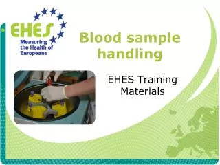
Whole Blood • Is placed into a container with an _____________________added to prevent clotting. • As soon as you obtain your sample, mix the blood with the anticoagulant by using a ____________________________ motion. • Vigorously shaking your sample can cause ______________, otherwise known as cell destruction.
_____________ • Defined: Are chemicals that prevent or delay the clotting process. • Choice of anticoagulant depends on _____________needed. • Sample must be _______________before use.
Anticoagulants cont’d • Samples not tested within _________of collection should be refrigerated. (Bring sample back to room temperature and re-mix before analysis. • ___________ blood should NEVER be frozen as the freezing/thawing process can _______ the blood cells.
Red Topped Tube • Red Topped Tube: Contains no ____________________. • Routinely used for ________________________. • Used for ________samples.
Serum Separator/Tiger Topped Tube • Tiger Topped (Striped) Tube/Serum Separator: • Contains no anti-coagulant. Has a yellowish “plug” of __________________________________that separates serum from plasma when spun. Used for ___________ samples. ***(Not used for therapeutic drug level monitoring.)
Lavender/Purple Topped Tubes • Lavender Topped Tube: Contains the anticoagulant __________ or Ethlenediamine tetraacetic acid. • Used for ____________________samples or __________samples. • Used for complete blood counts because it does not ____________________________. HOWEVER, an excess of anticoagulant in a sample may cause cells to __________and invalidate cell counts done on automated analyzers.
Grey Topped Tube: • Contains the anticoagulant _________________________. • Best for ___________________ preservation. • Interferes with many other tests performed on serum.
Blue Topped Tube • Contains the anticoagulant _______________________. • Commonly used in ___________________. • Na Citrate interferes with Na assays and many common serum tests.
Green Topped Tube • Contains the anticoagulant ________. • Can be used for most tests that require ____________samples. • Should never be used for differential blood film analysis because the anticoagulant interferes with the staining of the _______’s.
Hematology • Defined: ____________________________
Why is hematology important? • Evaluation of _________________ • Screening for _______ animals as a _______________. • ___________________ screening • _______-___________ monitoring
Hematopoiesis • Refers to the production of __________________________ and __________________. All blood cells arise from the same _______________________________________.
Blood Composition • Blood is composed of __________and __________ • Fluid portion is ~____% water
Packed Cell Volume • The PCV is measuring the percent of CELLS in a patient’s blood. • If the animal is dehydrated, the fluid portion of the blood will ____________. • Example: a PCV of 50% will give a sample that is 50% cells and 50% fluid. This means that a 10mL sample will yield 5mL of fluid. A PCV of 70% will yield 70% cells and only 30% fluid so a 10mL sample will only give 3mL of fluid.
PCV (Packed Cell Volume) • In a CBC, we determine the number of RBC’s in several different ways. The quickest and easiest is called the __________________, also referred to as the packed cell volume (PCV) • The PCV will tell you if the animal is ________or ________.
Normal PCV Values • Canine: _______%Feline: _______%Equine: _______%Bovine: _______%
Whole blood is collected in an ______________ (usually EDTA) and placed in a capillary tube. • Microhematocrit tubes should be filled to the designated line, with one end plugged with clay sealant.
Blood sample should be spun in a microhematocrit centrifuge for ______ minutes • Lay the tube in the centrifuge with the plugged end facing the ___________of the centrifuge. Make sure that a _______________is placed opposite or have another sample across from yours. • Cells are ________than the plasma and are compacted at the end of the tube that has the clay plug.
Plasma Evaluation • Plasma color and transparency may be helpful in determining a diagnosis and should be recorded in your findings. • Normal plasma is _____or a _____________color • Cloudy Plasma = ________ • Reddish tinge = _________ • Yellow = ________(indicates possible liver disease)
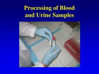
Concentration of total protein / total solids • Plasma protein concentrations estimated with a _____________and is an important component of the CBC in all species. • Plasma used to determine the TP/TS is collected by breaking the hematocrit tube just above the _________/_________interface. ↵
The plasma is allowed to flow onto the ______________________________________. (Blow gently through the open end of the hematocrit tube with the broken end of the tube over the prism of your refractometer.) • Hold the refractometer up to the light and record your findings. • Make sure to ____________your refractometer after each use!
Blood Films • The blood film is used to perform the ____________, estimate platelet numbers; and evaluate the _________________________________features of WBCs, RBCs and platelets. • _________________ smears are prepared by placing a small drop of blood on a clean glass microscope slide
Staining a slide • Always stain using the ________to _______stain. • Remember which side of your slide is up (clothes pins are marked “top”) • Rinse off from _____________side of slide • May _______________ to speed up process. (We will NOT be doing this!)
Performing the Differential Cell Count • This is where the different _____________________________________ are tallied separately. This can be done by a blood counting machine, or by hand. • To ________count the different cells, first you must make a ____________. Stain the slide once it is dry. • Using a cell counter you will tally a total of _____cells (this will make it easy to turn the numbers into a %)