Download
1 / 61
610 likes | 638 Views
Explore the conducting and respiratory portions, trachea and bronchial tree features, and alveolar wall structure explained by Prof. Ji-Cheng Li. Learn about nasal cavity, pharynx, larynx, trachea, bronchi, and lung anatomy in detail with illustrations.

E N D
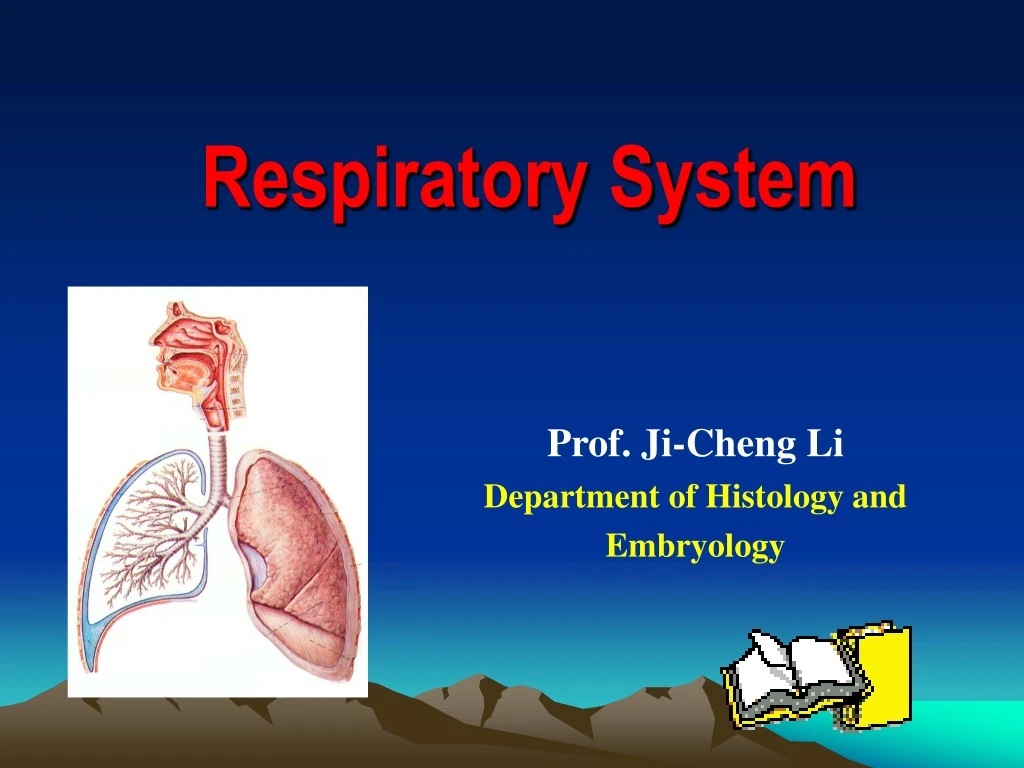
Respiratory System Prof. Ji-Cheng Li Department of Histology and Embryology
Objectives • Components of the conducting and respiratory portions of the respiratory system. • Wall structure and key distinguishing features of the trachea and the various segments of the bronchial tree. • Structure of the alveolar wall and components of the gas-blood barrier. • Structure of the type I, II alveolar cells.
Components Nasal cavity The pharynx (digestive system) The larynx The trachea The bronchi The lung
1.Nasal cavity (study by yourself) Ep: stratified squamous epi. LP: sebaceous and sweat gland vestibular region respiratory region olfactory region Ep: pseudostratified ciliated columnar epi. LP: vascular network Olfactory cells Supporting cells Basal cells Ep: olfactory epi. LP: serous gland (Bowman gland, olfactory gland)
2. Trachea and main bronchi Structure: • Mucosa • Submucosa • Adventitia
(1) Mucosa : Epithelium Pseudostratified ciliated columnar epithelium • Ciliated cell • Goblet cell • Brush cell • Basal cell • Small granule cell
Epithelium Figure 17-6: Ciliated respiratory epithelium
ciliated cell • with cilia • To provide a sweeping motion from the farthest reaches towards larynx
Goblet cell • to synthesize and secrete mucus • the secretion covers the epithelium surface
Brush cell -columnar, microvilli, -EM: RER, no granules -function: not very clear i. become into ciliated cell ii.receive sensory stimuli
Small granule cell (neuroendocrine cell) -EM: dense-core granules -Function: secret hormones to regulate contract of SM and secretion of gland i. 5-hydroxytryptamine(serotonin) ii. Calcitonin
Basal cell stem cell
(2) Submucosa LCT, containing mixed glands with diffuse LT and LN SIg A = secretory component (secreted by epithelum cell) + Ig A ( produced by plasma cell)
(3) Adventitia: cartilage ring: 16-20 “C ” shaped circular ligament: elastic fiber posterior part (membrane part): SM, elastic fiber, tracheal gland
3. Lung ---paired organ, located in thoracic cavity
General structure: ---capsule: visceral layer of pleura- serous membrane ---parenchyma: all branches of bronchi and alveoli ---interstitia
Conducting portion: bronchi →intrapulmonary bronchial tree (lobar bronchial tree, segmental bronchi and small bronchi) →small bronchi →bronchioles →terminal bronchioles • Function: inspire air (cleaned, moistened, warmed)
Respiratory portion respiratory bronchioles →alveolar duct →alveolar sac → alveoli Function: gas exchange
1) Conducting portion ① bronchi→small bronchi (fromlobar bronchi to small bronchi) • mucosa: Pseudostratified ciliated columnar epithelium -epithelium : thinner -goblet cell: number ↓ -lamina propria: thinner, -SM ↑ • submucosa:gland ↓ • adventitia:cartilage ↓
② bronchiole: • diameter< 1mm • pseudostratified ciliated columnar epithelium • goblet cell, Gland, cartilage↓ or disappear • smooth muscle ↑ • circular mucosa plica ↑
*pulmonary lobule: one bronchioles and its all branches and all alveoli cone or pyramidal-shape 0.1 cm in diameter
*Asthma • Allergy--"the epidemic of the 21st century". • Asthma is a serious disease that affects the lungs and the airways that deliver air to the lungs.
③ terminal bronchiole: Diameter < 0.5 mm ---goblet cell, gland, cartilage disappear ---SM: form a whole layer of circumferential SM ---Wall: • simple ciliated columnar epithelium two types of cells
i. ciliated cell ii. secreting cell: Clara cell non-ciliated and contain rich secretory granules (proteolytase) function: • dissolve the mucus • undifferentiated cell→ ciliated cell
Summary: Changes of conduction portion • The cartilages become irregular,and are smaller. • The amount of muscle in the bronchial wall increase. • Glands become fewer,and are absent in the bronchioles. • The epithelium become thinner.
bronchi →small bronchi bronchiole terminal bronchiole
2) respiratory portion ① respiratory bronchiole • simple columnar or cuboidal epithelium • smooth muscle less
② alveolar duct: ---wall: alveoli or alveolar sac opening simple cuboidal epithelium or squamous epithelium
③ alveolar sac: ---many alveoli open to it
④ alveoli: ---with opening alveolar sac 0.2mm in diameter, 300-400 million/per lung, total area: 70-80mm2 ---wall squamous epithelium
Respiratory bronchiole Alveolar duct Alveolar sac Alveoli
Epithelium of Alveoli • type I alveolar cells: • squamous, • cover 95% of the alveolar surface • type II alveolar cell • cuboidal • cover 5% of the alveolar surface
---type I alveolar cell: EM: • plasmalemmal vesicles • tight junction Function: constitute the blood-air barrier
---type II alveolar cell: secretory cells • contain osmiophilic multilamellar bodies • to release the surfactant to lower the surface tension • differentiate into type I alveolar cells.
EM: secreting granules: osmiophilic multilamellar body -0.1-1.0 um contains: phospholipid, glycosaminoglycan protein surfactant
alveolar septum: Ct , elastic and reticular fibers Fibroblast, macrophage, plasma cell, mast cell capillary: endothelium + basement membrane


