Download
1 / 12
120 likes | 366 Views
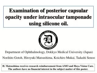
Examination of posterior capsular opacity under intraocular tamponade using silicone oil. Department of Ophthalmology, Dokkyo Medical University (Japan). Norihito Gotoh, Hiroyuki Matsushima, Koichiro Mukai, Tadashi Senoo.

E N D
Examination of posterior capsular opacity under intraocular tamponade using silicone oil. Department of Ophthalmology, Dokkyo Medical University (Japan) Norihito Gotoh, Hiroyuki Matsushima, Koichiro Mukai, Tadashi Senoo H. Matsushima receives research reimbursement from AMO and Hoya Vision Care. The authors have no financial interest in the subject matter of this poster.
Purpose Severe proliferative diabetic retinopathy Severe retinal disease patients sometimes undergo vitrectomy with intraocular tamponade using silicone oil. In these patients, posterior capsular opacity (PCO) developed markedly. However the mechanisms of PCO and effects of silicone oil to lens epithelial cells (LECs) are still unclear. In this study, we produced a novel cell culture system for analyze the effect of silicone oil to LECs experimentally. Posterior capsule fibrous opacity in silicone oil tamponade eye
Methods: Cell Culture Model Cultured LECs from rabbit (3.0x10³cells for HE-staining & 2.0x10⁴cells for immune-staining) with 10%FBS/MEM were embedded on upper chamber. The cell culture insert (12-well) has 1.0mm pores in the supporting PTE membrane for diffusion of medium between culture chambers. BSS group Intraocular irrigating solution (Balanced salt solution) was added in lower chamber. Air group The air group was blank in lower chamber. Silicone group Silicone oil was added in lower chamber. The LECs in cell culture insert were cultured during 7days for HE-staining and 4days for immune-staining. After the selected period, the LECs were fixed with10%formalin and were stained.
Methods:Histological and Immune-Histological Staining Hematoxylin-eosin (HE) staining The LECs were stained by hematoxylin-eosin to examine for the morphology of cells. The aspect ratio of LEC was calculated to assess the fibrosis of LEC. B B Aspect ratio of LEC (%) =x100 A A Immuno-histological staining The LECs were examined by immune-staining using anti-a-Smooth Muscle Actin (a-SMA) and anti-Transforming Growth Factor-b2 (TGF-b2) to study for fibrosis of the cells. The ratio of positive cell was calculated. Immune-staining positive cells Ratio of positive cell (%) = x100 Total nuclei
Results:Morphology of LEC by HE Staining 10x 20x 40x BSS group Air group Silicone group The morphology of LEC in Silicone and Air group was enlarged compared with LEC in BSS group. LECs in BSS group were kept in normal cubic structure.
Results: Aspect Ratio of LEC * % 100 80 60 40 20 0 BSS group Air group Silicone group The aspect ratio of LEC was 35.4% in silicone group, 65.8% in Air group, and 83.1% in BSS group. There are statistically significance (Tukey-Kramer method *P<0.05).
Results: Immune-Histological Staining for a-SMA 10x 20x 40x BSS group Air group Silicone group The LEC in Silicone group was higher expressed a-SMA than the LEC in Air and BSS group.
Results: Ratio of Positive Cell for a-SMA % * 100 * 80 60 40 20 0 BSS group Air group Silicone group The ratio of positive cell for a-SMA was 62.2% in silicone group, 28.1% in Air group, and 14.9% in BSS group. There are statistically significance (Tukey-Kramer method *P<0.05).
Results: Immune-Histological Staining for TGF-b2 10x 20x 40x BSS group Air group Silicone group The LEC in Silicone group was higher expressed TGF-b2 than the LEC in Air group. The LEC in BSS group was nothing TGF-b2 expression.
Results: Ratio of Positive Cell for TGF-b2 % * 50 40 30 20 10 0 BSS group Air group Silicone group The ratio of positive cell for TGF-b2 was 30.7% in silicone group and 5.4% in Air group. The LEC in BSS group was nothing TGF-b2 expression. There are statistically significance (Tukey-Kramer method *P<0.05).
Hypothesis Surviving LEC Fibroblast-like cell transform a-SMA(+) TGF-b2 Smads 3/4 ECM (extracellar matrix) nuclear translocation PCO EMT (posterior capsular opacity) Silicone oil (epithelial-mesenchymal transition) The silicone oil tamponade after vitrectomy may promote TGF-b2 from surviving LEC in lens capsule and transform from the LEC into fibroblast-like cell by EMT. The accretion of EMT and increasing of fibroblast-like cells may produce severe PCO.
Conclusions The intravitreous substituted materials have much effect on lens epithelial cell. The mechanisms of posterior capsular fibrous opacity after vitrectomy is indicated that the intravitreous materials, in particular silicone oil, undergo epithelial-mesenchymal transition.