Download
1 / 51
990 likes | 2.78k Views
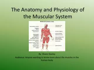
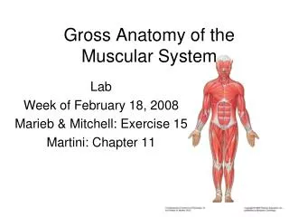
Anatomy of the Muscular System. Anatomy & Physiology. Skeletal Muscle Structure. Endomysium : connective tissue membrane that surrounds individual muscle fibers or cells Fascicles : groups of skeletal muscle fibers Perimysium : connective tissue membrane surrounding fascicles

E N D
Anatomy of the Muscular System Anatomy & Physiology
Skeletal Muscle Structure • Endomysium: connective tissue membrane that surrounds individual muscle fibers or cells • Fascicles: groups of skeletal muscle fibers • Perimysium: connective tissue membrane surrounding fascicles • Epimysium: connective tissue membrane around muscle as a whole
Attachment to bones • Tendons: strong touch cord • Aponeurosis: broad flat sheet of connective tissue
Fascia • Fibrous connective tissue surrounding the muscle organ & tendon
Attachment of Skeletal Muscles • Origin: point of attachment that doesn’t move when muscle contracts • Insertion: point of attachment that moves when muscles contract
Muscle Actions • Agonist (prime mover) • Antagonist • Synergist • Fixator
Agonist and antagonist • Agonist: muscle or group of muscles that that directly perform a specific movement • Antagonist: muscles that oppose a prime mover or agonist. Relaxed when agonist when contracts
Synergist & Fixator • Synergists: muscles that contract at the same time as the prime mover. They complement prime mover actions. • Fixator: generally function as joint stabilizers. Serve to maintain posture or balance.
How muscles are named • Location: biceps, brachii • Function: adductor, longus • Shape: trapezius • Direction of fibers: rectus, abdominus • Number of heads: biceps, brachii • Points of attachment: sternocleidomastoid • Size: gluteus, maximus
Muscles of facial expression Occipitofrontalis #6 Orbicularis oris #4 Orbicularis oculi #2 Zygomaticus #3 Buccinator Muscles of Mastication Massester #15 Temporalis #1 Muscles of the Head
Muscles of the Neck • Sternocleidomastoid #16 front view • Trapezius #6 back view
Muscles of the Thorax • Internal intercostals • External intercostals • Diaphragm
Muscles of the Abdominal Wall • External oblique #17 front • Internal oblique #9 front • Tranversus abdominus • Rectus abdominus #8 front
Biceps brachii • Flexes forearm • # 7 front view
Triceps brachii • Extends the lower arm • #8 back view
Pectoralis • #5 front view
Deltoids • #6 front view
Serratus anterior • #23 front view
Teres major • #12 back view
Pronator • # 24, front view
Bachioradialis • #25 front view
Flexor carpi radialis • #26 front view
Gluteus maximus #1, back view
Gluteus medius • #11 back view
Pectineus • #27 front view
Gracilis • #11 front view
Sartorius • #12 front view
Adductor magnus • #2 back view • #10 front view
Rectus femoris • #19 front view
Vastus lateralis • #20 front view
Vastus medialis • #21 front view
Biceps femoris • #3 back view
Semitendinosus • #4 back view
Peroneus • #22 front view
Tibialis anterior • #13 front view
Soleus • #13 back view
Gastrocnemius • #5 back view
Image Citations • Slide 3: Structure of skeletal muscle, 11/5/06, http://www.people.eku.edu/ritchisong/301notes3.htm • Slide 4, 8, 13: • Slide 6: muscle, 11/5/06, http://www.octc.kctcs.edu/gcaplan/anat/Notes/API%20Notes%20J%20Anatomy%20of%20a%20Muscle.htm • Slide 17: Biceps brachii, 11/5/06, http://homepages.utoledo.edu/smcgreg2/ANTMUSCLE/sld004.htm • Slide 18: Triceps brachii, 11/5/06, http://de.wikipedia.org/wiki/Triceps_brachii • Slide 19: Pectoralis major, 11/5/06, http://www.exrx.net/Muscles/PectoralisSternal.html
Image Citations • Slide 20: Deltoids, 11/5/06, http://www.cartage.org.lb/en/kids/science/Biology%20Cells/Muscle/Kids%20Muscles/Shoulder/Shoulder.htm • Slide 21: Serratus anterior, 11/5/06, http://pl.wikipedia.org/wiki/Mi%C4%99sie%C5%84_z%C4%99baty_przedni • Slide 22: Teres major, 11/5/06, http://www.abcbodybuilding.com/anatomy/shouldersanatomy1.htm • Slide 23: Brachialis, 11/5/06, http://www.letempledelaforme.com/anatomie/anatomie/brachial_anterieur.htm • Slide 24: Pronator teres, 11/5/06, http://www.rad.washington.edu/atlas/pronatorteres.html
Image Citations • Slide 25 & 26: Brachioradialis, 11/5/06, http://www.octc.kctcs.edu/gcaplan/anat/Notes/API%20Notes%20J%20%20Muscles%20of%20the%20Arm.htm • Slide 27: Flexor carpi radialis, 11/5/06, http://www.octc.kctcs.edu/gcaplan/anat/Notes/API%20Notes%20J%20%20Muscles%20of%20the%20Arm.htm • Slide 28: Gluteal muscles, 11/5/06, http://www.audreysmassage.com/featurepage/gluteal.html • Slide 29: Gluteus medius, 11/5/06, http://www.audreysmassage.com/featurepage/gluteal.html • Slide 30: Diagram of pectineus m, 11/5/06, http://summit.stanford.edu/ourwork/PROJECTS/LUCY/lucywebsite/pectineus1.html
Image Citations • Slide 31: Anterior hip muscles, 11/5/06, http://www.answers.com/topic/gracilis-muscle • Slide 32: Sartorius, 11/5/06, http://www.sportandsupport.nl/sartorius.html • Slide 33: Adductor magnus and longus, 11/5/06, http://courses.washington.edu/hubio553/atlas/205.html • Slide 34: Rectus femoris, 11/5/06, http://www.sante.cc/electro/dossiers/quadrifemo/fig01.htm • Slide 35: Vastus lateralis, 11/5/06, http://myphlip.pearsoncmg.com/altproducts/drugguide/ab2page.cfm?vbcid=6645&vid=1110 • Slide 36: Vastus medialis, 11/5/06, http://www.sante.cc/electro/dossiers/quadrifemo/fig03.htm
Image Citations • Slide 37: Vastus intermedius, 11/5/06, http://www.sante.cc/electro/dossiers/quadrifemo/fig04.htm • Slide 38: Biceps femoris, 11/5/06, http://www.gomotion.dk/pe1.asp?p_id=171 • Slide 39: Hamstring muscles, 11/5/06, http://www.gla.ac.uk/ibls/fab/tutorial/anatomy/thight.html • Slide 40: Semitendinosus, 11/5/06, http://www.octc.kctcs.edu/gcaplan/anat/Notes/API%20Notes%20J%20%20Muscles%20of%20the%20Leg.htm • Slide 41: Peroneus, 11/5/06, http://biking.taiiku.tsukuba.ac.jp/~takai/Anatomy/e-anatomy/GR/peroneus.l.GIF • Slide 42: diagram of tibialis anterior, 11/5/06, http://vancouvermassage.ca/articles/spot-03.php