The Nervous System:
240 likes | 530 Views
The Nervous System:. Tissues and the Spinal Cord. Nervous & Endocrine Systems. Compare the mode of communication in these two systems. (Complete the worksheet on page 123 in your course packet. Organization of the Nervous System.

The Nervous System:
E N D
Presentation Transcript
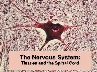
The Nervous System: Tissues and the Spinal Cord
Nervous & Endocrine Systems Compare the mode of communication in these two systems. (Complete the worksheet on page 123 in your course packet.
Organization of the Nervous System In this flow chart, what do the components in blue have in common? Explain why the Enteric Nervous System is referred to as the “little brain”. Where are the Autonomic sensory receptors located?
Basic Tasks of the Nervous System Sensory Input: Receptors monitor both external and internal environments. Integration: Process the information (at synapses) and often integrate it with stored information. Motor output: If necessary, signal effector organs to make an appropriate response. How is this similar to the normal function of the endocrine system? How is it different? Describe the receptor, control center, and effector in several neural reflexes.
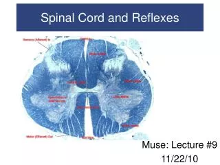
Basic Structure of a Reflex Arc Review the lab exercise on reflexes in your packet, and complete the worksheet that follows.
Multipolar neuron Are dendrites always shorter than axons? Can the neuron processes that conduct information toward the cell body also be myelinated?
Review: Structural Classificationof Neurons Compare the possible functions of these neuron types. Give one location where these neurons would be found in the body.
Gray and White Matter For practice, label the parts of the brain and spinal cord visible in these sections. What structures are found predominately in gray matter? …in white matter? Why do you think white matter surrounds gray matter in the spinal cord?
Neuroglia of the CNS • Astrocytes • Oligodendrocytes • Microglia • Ependyma Which neuroglia have a protective function? Which neuroglia provide myelination in the CNS? Which neuroglia are involved in transport? Name the cell type that provides myelination in the PNS. How are oligodendrocytes and Schwann cells different?
Astrocytes This picture illustrates the relationship between astrocytes and blood vessels. The dark 'star-like' figures are the astrocytes. http://blustein.tripod.com/Astrocytes/astrocytes.htm
Organization of Neurogliain the CNS What differences between gray matter and white matter are visible in this figure? From which embryonic germ layer do these neuroglia originate? What is the blood-brain barrier?
Review of Chemical Synapses Compare with the structure and function of an electrical synapse. How does the action differ between an excitatory neurotransmitter and an inhibitory one?
Propagation of an Action Potential Why is the resting potential a negative number? Refractory Period What is the significance of the refractory period?
Saltatory Conduction in Myelinated Axons Action potentials jump from node to node without depolarizing the region under the myelin sheath - called saltatory conduction. What cells myelinate fibers in the CNS? In addition to the presence of a myelin sheath, what else may increase the speed of conduction along a neuron process? Fig. 48.11
Structure of Peripheral Nerves Are nerves considered organs of the nervous system? Why is there such a large c.t. component to nerves?
Regeneration of Peripheral Nerve Fibers (1) How might this injury occur? Is this a sensory neuron, interneuron, or motor neuron? (What function would be lost?)
Regeneration of Fibers in the PNS (2) What is this component of the nerve?
Regeneration of Fibers in the PNS (3) What is a neuroma?
Cranial and Spinal nerves of the PNS Label the nerves in these figures. Which nerves are classified as sensory?…motor?…mixed?
Embryonic Origin of the Central Nervous System Use this figure to help interpret the flow chart in your course packet.
Structure and Function of theSpinal Cord There are 31 segments to the spinal cord; each segment giving rise to a pair of ____________. Name the two main functions of the spinal cord. Why are there cervical and lumbar enlargements?