Download
1 / 35
360 likes | 396 Views
Learn about head injuries, including skull fractures, brain injury, and concussions. Explore the pathophysiology, diagnosis, and medical management of head trauma. Discover the clinical manifestations and residual effects of brain injuries.

E N D
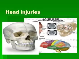
Head Injury • Most common cause of death due to trauma • Includes injury to scalp, skull or brain • Highest between 15 to 24 • Best approach to head injury is prevention
Pathophysiology • Not all brain damage occurs at the moement of impact • Damage – two forms Primaryinjury : initial damage at the time of the traumatic event – includes contusions, lacerations and torn blood vesselsfrom impact, acceleration / deceleration or foreign objects penetraion Secondaryinjury : evolves over the ensuing hours and days after the initial injury and is due primarily to brain swelling or ongoing bleeding
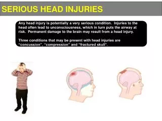
Rigid skull – swelling – the space cannot hold more – ICP increases – contents of the skull get displaced – downward or lateral – pressure leads to restriction of blood flow – brain death – herniation
Scalp Injuries • Abrasion • Contusion • Laceration • Haematoma (eg. Subgaleal haematoma) Head injuries are portals of intracranial infections and hence should be irrigated well before suturing
Skull Fractures • With or without damage to the brain • Persistent localized pain usually suggests a fracture • Fractures of the cranial vault may or may ntot produce swelling the region of the fracture; therefore, and X-ray is needed • Fracture of the base of skull may traversethe paranasalsinusthe middleear located in the temporal bonehence bledding from the nose, pharynx, or ears and blood may appear under the conjuctivabruising over mastoid – Battle’ssign
Skull Fractures • CSF rhinorrhoea, CSF otorrhoea • A blood stain surrounded by a yellowish stain – halo sign in bed linens or the head dresing and is highly suggestive of a CSF leak. Drainage of CSF – meningeal infection • Bloody CSF suggests a brain laceration or contusion
Diagnosis • CT scan • MRI when the patient is stable enough to undergo this longer diagnostic study • Cerebral angiography – to identify supratentorial, extracerebral and intracerebral haematomas and cerebral contusions. • Lateral and anteroposterior views of the skull
Medical Management • Nondepressed skull fractures : no surgical treatment; close observation • Contaminated and deforming depressed fractures need surgery • Large defects in the skull repaired with bone or artificial grafts • Penetrating wounds : debridement and removal of foreign bodies and devitalized tissue • Antibiotic • Blood component therapy if indicated • Base of skull fractures : opening into sinuses, nose, ear, pharynx – sterile cotton is placed to keep the cavity clean and to collect the draining fluid • Caution against sneezing, coughing and blowing nose • Persistent CSF rhinorrhoea or otorrhoea : surgical intervention
Clinical Manifestations of Brain Jnjury • Altered level of consciousness • Confusion • Pupillary abnormalities (changes in shape, size and response to light • Altered or absent gag reflex • Absent corneal reflex • Sudden onset of neurologic deficits • Changes in vital signs (altered respiratory pattern) hyper tension, bradycardia, tachycardia, hypothermia or hyperthermia • Vision and hearing impairment • Sensory dysfunction • Spasticity • Headache • Vertigo • Movement disorders • seizures
Brain Injury • Closed brain injury • Open brain injury Concussion : temporary loss of neurologic function with no apparent structural damage. A period of unconsciousness lasting from a few seconds to a few minutes. The injury may be mild and may cause only dizziness or severe enough to cause complete loss of consciousness for a time. Frontal lobe : bizarre irrational behaviour Temporal lobe : temporary amnesia or disorientation
Patient may be observed for 24 hours Observe for postconcussion syndrome : headache, dizziness, lethargy, iritability and anxiety • Normal activities should be started slowly • The family instructed to look for :difficulty in awakeningdifficulty in speakingconfusionsevere headache vomiting weakness of one side of the body
Residual effects of concussion : headache Lethargy personality changes changes in behaviour Attention deficits Difficulty with memory Disruption in work habits Gerontologic considerations : even with minor injuries the elderly may suffer more serious damage. Any elderly patient with behavioural disturbances or confusion should be assessed for head injury, because unrecognized “minor” head trauma may account for behavioural and confusional episodes
Contusions of right laeral temporal lobe, contusions anterior to frontal horns of lateral ventricles, extensive contusions along corpus callosum, left frontal lobe epidural haematoma with mass effect on left frontal cortex, fluid in maxillary sinuses
Contusion Brain bruised Surface haemorhage may be present Unconsciousness Motionless Faint pulse Shallow respirations Cool pale skin Involuntary evacuation of bowels and bladder Patient may be aroused with effort but soon slips BP and temperature are subnormal
In general patients with severe brain injury who have abnormal motor function, abnormal eye movements and elevated ICP have poor outcomes.- brain damage, disability or death. • Conversely the patient may recover consciousness but pass into a stage of cerebral irritability. In this stage, the patient is conscious and easily disturbed by any form of stimulation such as noises, light and voices. May become hyperactive at times • Then gradually returns to normal months later • Residual headache and vertigo are common • Impaired mental function or seizures may occur as a result of irreparable cerebral damage
Diffuse axonal injury • Involving cerebral hemispheres, corpus callosum, and brain stem • Axonal swelling and disconnection • No lucid intervals • Immediate coma • Decorticate posture • Decerebrate posture • Diagnosis by clinical signs in conjunction with a CT scan or MRI.
Intracranial Haemorrhage • Haematomas within the cranial vault are the most serious brain injuries • Epidural • Subdural • Intracerebral • Major symptoms delayed until distortion of the brain or ICP increase • Hematoma – compression – ischaemia • Rapid hge more dangerous even if small and slow he even if large may allow compensatory phenomena
Epidural Haematoma(extradural haematoma or hemorrhage) • An extreme emergency • Blood in the epdural space • Skull fracture may be + • Middle meningeal artery ruptures • Expanding haematoma causes symptoms • Initially a momentary loss of consciousness at the time of injury • An interval of apparent recovery may follow (lucid interval) • Then ICP increases • Suddenly signs of cpmpression appear : deterioration of consciousness and signs of focal neuroloogic deficits such as dilation and fixation of a pupil or paralysis of an extremity) and the patient deteriorates rapidly. • Respiratory arrest can occur within minutes • Burr hole, decompress brain. Craniotomy evacuate clot control bleeding, drain kept
Subdural Haematoma • Between the dura and the brain • Trauma, coagulopathies or rupture of an aneurysm • Usually venous; involving small vessels that bridge the subdural space. • Acute, subacute or chronic Acute and subacute subdural haematoma • Major head injury : contusion or lacetation • Clinical symptoms develop over 24 to 48 hrs. • Rapidly expanding mass coma, increasing blood pressure decreasing heart rate and slowing respiratory rate * • Immediate intervention is needed
Chronic Subdural Haematoma • Can develop from seemingly minor head injuries • Most frequent in the elderly • Brain atrophy • Brain shifts abnormally even on small impact • The veins which bridge the brain substance and the skull snap bleed – bleeding is less profuse • Symptoms delayed by weeks to months • Injury forgotten • May be mistaken for a stroke • The symptoms fluctuate • Headaches come and go • Alternating focal neurological signs • Personality changes • Mental deterioration • Focal seizures • Patient may be labelled a neurotic or psychotic • Burr holes or craniotomy: evacuate clot or mass of ossified or calcified clot
Intracerebral Haemorrhage or Haematoma • Bleeding into the substance of the brain • Missile injuries or bullet wounds stab injuries • Systemic hypertension • Rupture of a vessel or aneurysm • Vascular anomalies • Intracranial tumours • Bleeding in leukemia, haemophilia, aplastic anemias, thrombocytopenic purpuras, anticoagulant therapy • Onset insidious • Neurologic deficit followe by headache
Intracerebral Haemorrhage or Haematoma - management • Control of ICP • Fluids, electrolytes • Antihypertensives • Surgical : craniotomy or craniectomy and removal of clot and control haemorhage if possible. If very deep and inaccessible or uncircumscribed nothing could be done
Management of Brain Injuries • Prevent secondary injury due to increasing ICP and maintain cerebral oxygenation • Evacuate clot • Debride wound • Elevate depressed fractures of the skull • Suture scalp lacerations • Monitor ICP • Elevate head end of bed • Maintain blood volume • CSF may be drained by insertion of a drain
Supportive Measures • Ventilatory support • Siezure prevention • Fluid and electrolyte maintenance • Nutritional support • Pain and anxiety managed – • Benzodiazepines for fits • Nasogastric tube and aspiration to prevent aspiration common in the first few hours
Brain Death • Brain death act • Death will be determined with accepted medical standards and that death will indicate irreversible loss jof all brain function • The patient has no neurologic activity upon clinical examination ; adjunctive tests such as EEG and cerebral blood flow studies are often used to confirm brain death • Many of these patients are potential organ donors and the family may be informed.
Glasgow Coma Scale Used to assess patient’s response to stimuli Scores rang from 3 (coma) to 15 (normal)
Nursing Interventions • Monitor for declining neurologic functionLOC regularly assessedlook for vital signs ; bradypnoea, bradycardia, increase in BPmotor functionother neurologic signs : spontaneos eye opening, unilaterally dilated and poorly responding pupil (developing haematoma), pressure on third nerve due to shift of brain, anosmia, aphasia • Maintain airway • Monitor fluid and electrolyte balance • Promote adequate nutrition • Prevent injury
Maintain body temperature • Maintain skin integrity • Improve cognitive functioning • Preventing sleep pattern disturbance • Supporting family coping • Monitoring and managing potential complicationsdecreased cerebral perfusioncerebral oedema and herniationimpaired oxygenation and ventilationimpaired fluid, electrolyte and nutritional balancepost-traumatic seizures • Promoting home and community-based careteaching patients self-carecontinuing care
Controlling ICP in Brain Injury • Elevate the head of ved 30 degrees • Maintain the patient’s head and neck in neutral alignment (no twisting) • Initiate measures to prevent the Valsalva maneuver ( eg. Stool softeners) • Maintain normal body temperature • Adminster O2 to maintain PaO2>90mmHg • Maintain fluid balance with normal saline solution • Avoid noxious stimuli (eg. Excessive suctioning, painful procedures) • Administer sedation to reduce agitation • Maintain cerebral perfusion pressure > 70 mm Hg