Download

1 / 126
1.36k likes | 2.13k Views
osteoarthritis. Dr.A.Noori Rheumatologist www,arrh.ir. Introduction. Osteoarthritis is a common disorder of synovial joints.

E N D
osteoarthritis Dr.A.Noori Rheumatologist www,arrh.ir
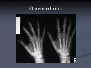
Introduction • Osteoarthritis is a common disorder of synovial joints. • Strongly age-related, being less common before 40 years,but rising in frequency with age, such that most people older than 70 years have radiological evidence of osteoarthritis in some joints.
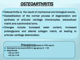
Epidemiology • Most common joint disease in human • Most frequent rheumatic compliant • Common cause of disability in elderly • Over 20 million affected in U.S. • About 12% :age>60 y • 6%:age>30 y
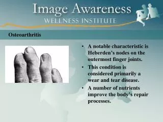
Nodal OA involving DIP and PIP joints is more common in women and their first degree female relatives • OA of knee is more common in African American women
Age Gender( female) Race Genetic factors Obesity sport Repetitive stress & joint overload Prior inflammatory joint disease Congenital /developmental defect Major joint trauma Metabolic /endocrine disorder muscle weakness Risk factors
OA – Risk Factors Age • Age is the strongest risk factor for OA. Although OA can start in young adulthood, if you are over 45 years old, you are at higher risk. Female gender • In general, arthritis occurs more frequently in women than in men. after age 45, OA is more common in women. OA of the hand is particularly common among women. Joint alignment • People with joints that move or fit together incorrectly, such as bow legs, a dislocated hip, or double-jointedness, are more likely to develop OA in those joints.
OA – Risk Factors Hereditary gene defect • A defect in one of thegenes responsible for the cartilage component collagen can cause deterioration of cartilage. Joint injury or overuse caused by physical labor or sports • Traumatic injury (ex. Ligament or meniscal tears) to the knee or hip increases your risk for developing OA in these joints. Joints that are used repeatedly in certain jobs may be more likely to develop OA because of injury or overuse. Obesity • Being overweight during midlife or the later years is among the strongest risk factors for OA of the knee.
Risk factors you cannot change • Family history of disease
Risk factors you cannot change • Family history of disease • Increasing age
Risk factors you cannot change • Family history of disease • Increasing age • Being female
Risk factors you can change • Overuse of the joint
Risk factors you can change • Overuse of the joint • Major injury
50% decrease in OA with with 11# wt loss Larger effect in women Strong Risk Factor for OA Obesity
Risk factors you can change • Overuse of the joint • Major injury • Overweight • Muscle weakness
Joint • EPIPHYSEAL BONE • CARTILAGE • SYNOVIAL MEMBRANE • CAPSULE • LIGAMENTS • MUSCLE & TENDONS • BURSAE
Cartilage • Function : • Reduce friction in the joints • Lubrecin • Water cushion • Absorb the shock associated with locomotion
Cartilage • Consist of : • Water :70% • Type II collagen • Proteoglycan : • Aggrecan Sub Unit • Core Protein • Glycosaminoglycans, Link Protein • Hyaluronic Acid • Chondrocyte
Cartilage • Layer: • Superficial • Intermediate • Deep • Tidemark • Calcified cartilage • Subchondral
IX XI II Collagen • Compressible • Elasticity • Self- lubrication
Cartilage metabolism • Cartilage is metabolically active • Synthesis matrix • Destruct matrix
Plasmine TPA factor Elastase Serine proteases Collagenases Gelatinases Stromelysines Metloproteinases (MMP) Proteinases Systeine proteases Catepsines
Cartilage remodeling Synthetic activities Degradative activities
synthesis activity = degradative activity h o m o s t a s I s Matrix degradation Matrix synthesis CHONDROCYTE
synthesis activity = degradative activity h o m o s t a s I s Matrix degradation Matrix synthesis collagen proteinases proteoglican CHONDROCYTE synoviocyte
synthesis activity = degradative activity h o m o s t a s I s Matrix degradation Matrix synthesis collagen proteinases proteoglican activation CHONDROCYTE IL-1 , TNF synoviocyte inflammation
synthesis activity = degradative activity h o m o s t a s I s Matrix degradation Matrix synthesis collagen proteinases proteoglican activation CHONDROCYTE NO synthase up regulation IL-1 , TNF IL-1 , TNF NO synoviocyte inflammation
synthesis activity = degradative activity h o m o s t a s I s Matrix degradation Matrix synthesis collagen proteinases proteoglican apoptosis activation CHONDROCYTE NO synthase Up regulation IL-1 , TNF IL-1 , TNF NO synoviocyte inflammation
synthesis activity = degradative activity h o m o s t a s I s Matrix synthesis Matrix degradation collagen proteinases proteoglican Inferior quality of matrix CHONDROCYTE Collagen I Collagen III differentiation TGF IGF bone
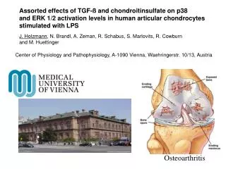
OA • OA is a disease of joints that affects all of the weight-bearing components of the joint: • Articular cartilage • Menisci • Bone
Erosion, detachment of fragments of cartilage subchondralmicrocyst & sclerosis Macroscopic Change
Core Protein-Aggrecan Hyaluronic Acid Link Glycoprotein Chondroitin Sulfate Chain Articular Cartilage Glycosaminoglycan
Bone change • Overactivity of Osteoblast Growth factor Subchondral sclerosis • Cartilage destruction • Bone hypertrophy (subchondral • Sclerosis ) • Osteophyte formation TGFβ, ILGF¹