Download
1 / 38
390 likes | 570 Views
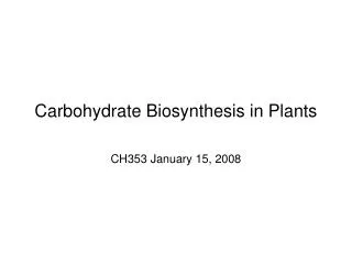
Current updates and evolving concepts on Cellulose Biosynthesis in plants. Debarati Basu Cell Wall Seminar. Outline. What genes involved in cellulose biosynthesis? Which compartment is involved in cellulose biosynthesis?

E N D
Current updates and evolving concepts on Cellulose Biosynthesis in plants Debarati Basu Cell Wall Seminar
Outline • What genes involved in cellulose biosynthesis? • Which compartment is involved in cellulose biosynthesis? • What are the protein components of the cellulose syntheses machinery and how they are coupled in the cytoskeleton? • What is the first committed step in cellulose polymerization? • How trafficking of cellulose synthase occurs? • What are the approaches in studying cellulose synthase?
Aaron H. Liepman, Raymond Wightman, Naomi Geshi, Simon R. Turner and Henrik Vibe Scheller (2010) Arabidopsis – a powerful model system for plant cell wall research The Plant Journal 61, 1107–1121.
The CESA proteins that make up the CSC responsible for primary cell wall formation consist of CESA1 and CESA3, together with some combination of CESA2, CESA5, CESA6, or CESA9.ACesA2 and CesA5, which have been reported to bepartiallyredundant with CesA6, may compete for the same position in the enzyme complex • Cellulose biosynthesis at the secondary wall requires CESA4, CESA7, and CESA8. Illustration of the structure of CESA3, a typical CESA protein. Chris Somerville (2006) Cellulose synthesis in higher plants Annu. Rev. Cell Dev. Biol. 22:53–78
Schematic model of cellulose synthesis Chris Somerville (2006) Cellulose synthesis in higher plants Annu. Rev. Cell Dev. Biol. 22:53–78
Cellulose synthase complex • Plasma membrane bound complex. • Arosette exhibits a typical diameter in the range 25–30 nm and consists of 6 globular structures arranged with a six-fold symmetry. • Eight transmembrane helices that anchor the proteins in the plasma membrane • The conserved D,D,D,QXXRW motif involved in the catalytic event. • Other accessory proteins might be involved along with the microtubules.
Cellulose synthase complex parameters EF Crowell, M Gonneau, Y-DStierhof, H Ho¨ fte and S vernhettes (2010) Regulated trafficking of cellulose synthases Current Opinion In Plant Biology 13: 1-6.
Figure 1. Hypothetical model for the biosynthesis of cellulose in higher plants. GeaGuerriero, Johanna Fugelstad and Vincent Bulone (2010) What Do We Really Know about Cellulose Biosynthesis in Higher Plants? Journal of Integrative Plant Biology 52 161–175.
Cellulose synthase trafficking • Involvement of cortical microtubules in the movement of the cellulose synthasecomplex in the plasma membrane. • Recent studies have shown SmaCCs/MASCs (small CESA compartments / microtubule associated cellulose synthase compartments ) are highly dynamic compartments that appear to play key roles both as intracellular stores of the CSC and in its delivery to the plasma membrane.
Overview of CSC intracellular trafficking Raymond Wightman and Simon Turner (2010) plant physiology 153 427–432.
Assembly of the CESA subunits EF Crowell, M Gonneau, Y-DStierhof, H Ho¨ fte and S vernhettes (2010) Regulated trafficking of cellulose synthases Current Opinion In Plant Biology 13: 1-6.
Cellulose as cellulosic biofuel Lignin biosynthesis
Link between lignin and cellulose biosynthesis The effects of downregulation of Pt4CL1 expression on Pt4CL1 activity and lignin accumulation in transgenic aspen. Wen-Jing Hu, Scott A. Harding, Jrhau Lung, Jacqueline L. Popko, John Ralph, Douglas D. Stokke, Chung-Jui Tsai, and Vincent L. Chiang (1999) Repression of lignin biosynthesis promotes cellulose accumulation and growth in transgenic trees Nature Biotechnology 17 808-812.
Enhanced growth in transgenic aspen Wen-Jing Hu, Scott A. Harding, Jrhau Lung, Jacqueline L. Popko, John Ralph, Douglas D. Stokke, Chung-Jui Tsai, and Vincent L. Chiang (1999) Repression of lignin biosynthesis promotes cellulose accumulation and growth in transgenic trees Nature Biotechnology 17 808-812.
Sucrose metabolism and cellulose biosynthesis Haigler, C.H., M. Ivanova-Datcheva, P. S. Hogan, V. V. Salnikov, S. Hwang, L. K. Martin, and Delmer, D.P. (2001) Carbon partitioning to cellulose synthesis. Plant Molecular Biology47: 29-51.
Sucrose synthase as a component of the catalytic unit of Cellulose synthase A C B (A) The deduced amino acid sequence of sucrose synthase in Azuki bean.(B) immune blotting using antibodies raised against mung bean sucrose synthase. (C) Cellulose synthesis by immunoprecipitate preparation after incubation with UDP-glucose.
Sucrose synthase as a component of the catalytic unit of Cellulose synthase E D (D) Ultracel YM-3 membrane-retained products in (C) after treatment with β -1,4 or β -1,3-glucanase. (E) Membrane-retained cellulose using either UDP-[ 14 C]glucose (UDPG) or [ 14 C]sucrose plus UDP (sucrose/UDP) as substrates determined by incorporated glucose. Satoshi Fujii , Takahisa Hayashi and Koichi Mizuno (2010) Sucrose Synthase is an Integral Component of the Cellulose Synthesis Machinery Plant Cell Physiol. 51: 294–301
SG serves as primer for elongation of β -1,4-glucan chains LiangcaiPeng, Yasushi Kawagoe, Pat Hogan, and Deborah Delmer (2002 )Sitosterol-β-glucoside as Primer for Cellulose Synthesis in Plants 295 147 – 150.
Methods in studying cellulose biosynthesis • Genetic approaches Reverse genetics and mutant analysis • Microscopy • Live cell imaging • GC-MS- cellulose content analysis and linkage analysis.
Approaches in studying cellulose biosynthesis • Live cell imaging Live cell imaging of the CSC. The images are taken of YFP-CESA6 fusion within the epidermis of a cotyledon petiole cell (A) and pavement cells (B) and characteristic ring-like appearance in Golgi (C). Raymond Wightman and Simon Turner (2010) plant physiology 153 427–432.
Unanswered Questions • Process of assembly of individual β-(1→4)-glucan chains as microfibrilsis poorly understood. • The direct physical association of sucrose synthase with the cellulose synthasemachinery has not yet been demonstrated. • The role of sitosterol- β-glucosideas a primer for cellulose biosynthesis remains to be firmly demonstrated in vivo. • The mechanism of translocation of the cellulose chains across the plasma membrane has yet to be elucidated.
References • Aaron H. Liepman, Raymond Wightman, Naomi Geshi, Simon R. Turner and Henrik Vibe Scheller (2010) Arabidopsis – a powerful model system for plant cell wall research The Plant Journal 61, 1107–1121. • EF Crowell, M Gonneau, Y-DStierhof, H Ho¨ fte and S vernhettes (2010) Regulated trafficking of cellulose synthases Current Opinion In Plant Biology 13: 1-6. • GeaGuerriero, Johanna Fugelstad and Vincent BuloneWhat Do We Really Know about Cellulose Biosynthesis in Higher Plants? Journal of Integrative Plant Biology 2010, 52 : 161–175. • Haigler, C.H., M. Ivanova-Datcheva, P. S. Hogan, V. V. Salnikov, S. Hwang, L. K. Martin, and Delmer, D.P. (2001) Carbon partitioning to cellulose synthesis. Plant Molecular Biology47: 29-51. • Raymond Wightman and Simon Turner (2010) Trafficking of the Plant Cellulose SynthaseComplex Plant Physiology, 153:427-432 . • Satoshi Fujii , Takahisa Hayashi and Koichi Mizuno (2010) Sucrose Synthase is an Integral Component of the Cellulose Synthesis Machinery Plant Cell Physiol. 51: 294–301. • Somerville C (2006) Cellulose synthesis in higher plants. Annu Rev Cell Dev Biol22: 53–78. • Wen-Jing Hu, Scott A. Harding, Jrhau Lung, Jacqueline L. Popko, John Ralph, Douglas D. Stokke, Chung-Jui Tsai, and Vincent L. Chiang (1999) Repression of lignin biosynthesis promotes cellulose accumulation and growth in transgenic trees Nature Biotechnology 17 808-812.
Identification of a cellulose synthase-associated protein required for cellulose biosynthesis Ying Gu, Nick Kaplinsky, Martin Bringmann, Alex Cobb, Andrew Carrolla, ArunSampathkumar, Tobias I. Baskin, StaffanPersson, and Chris R. Somerville. PNAS 2010 107(29):12866-12871.
Fig. 1. Identification of CSI1. (A) Schematic representation of CESA and CSI1 proteins. (B) CSI1 interacts with three primary CESA proteins in yeast.
Interactive model of CSI Truncated coexpression network for primary wall cellulose-related genes using the AraGenNet
Phylogeny of CSI protein in land plants. Full-length CSI-like sequences were identified in GenBank using BLASTP and aligned using ClustalW.
GUS assay Promoter GUS analysis of CSI1::GUS (D, F, and H) and CESA3::GUS (E, G, and I
Expression pattern of the CSI1 genes assessed through GUS Assay
Mutant Analysis Fig. 2. Schematic representation of six T-DNA insertion sites in csi1. Morphology of 4-d-old dark grown seedlings: (Left to Right) Col-0 (wild-type) and csi1-1, csi1-2, csi1-3, csi1-4, csi1-5, and csi1-6 mutants.
Mutant analysis C F D Determination of Hypocotyl length (C) and growth rate (D) of dark-grown wild-type (Col-0) plants and csi1-3, csi1-6, and prc1-1 mutants. (E) SEM of dark-grown hypocotyls in wild-type plants and csi1 mutants: (Left to Right) Arabidopsis thaliana Columbia (Col-0), csi1-3, and csi1-6 mutants. (F) Cellulose content estimation.
Morphology of csi1 mutants. (A) RT-PCR analysis of CSI1 mRNA expression in various transfer DNA (T-DNA) insertion lines. (B-K) Phenotypic analysis of the mutants.
CSI1 is localized to CESA-like particles in dark-grown hypocotyls cells. Fig. 3. (A–D) Optical sections of epidermal cells in 3-d-old dark-grown hypocotyls expressing RFP-CSI1 (A and C) and YFP-CESA6 (B and D).(E) Plot of RFP-CSI1 particle velocity vs. (F) Histogram of measured RFP-CSI1 particle velocities
Localization of GFP (G–I) Localization of GFP-CESA3 (G), RFP-CSI1 (H), and merge (I)
YFP-CESA6 dynamics are altered in csi1-3 mutants Fig. 4. YFP-CESA6 localization in dark-grown hypocotyls cells is shown in wild-type plants (A and B) and csi1-3 mutants (C and D).(E) Histogram of measured particle velocities.
Movie • http://www.pnas.org/content/suppl/2010/07/01/1007092107.DCSupplemental/sm01 • http://www.pnas.org/content/suppl/2010/07/01/1007092107.DCSupplemental/sm02.mov
Polarized light analysis of csi1 mutants. Polarized-light micrographs of (A) wild-type and (B) csi1-1-mutant roots. (C) Quantification of retardance and azimuth
Conclusion • The CSI1 protein is the first non-CESA protein associated with primary CESA complexes not in the secondary cell wall. • CSI1 colocalizes with primary CESA complexes, and csi1 mutations affect the distribution and movement of CESA complexes. • The csi1 mutations appeared to decrease the degree to which cellulose microfibrils are coaligned.