Download
1 / 51
510 likes | 645 Views
FOOT & ANKLE. MOHAN LAL Consultant Orthopaedic & Foot/Ankle Surgeon Surrey & Sussex NHS Trust Spire Gatwick Park Hospital North Downs Hospital. Presentation by Chandar Lal. SUBCATEGORIES. FOOT AND ANKLE EXAMINATION COMMON FOOT DISORDERS ANKLE DISORDERS TENDON DISORDERS.

E N D
FOOT & ANKLE MOHAN LAL Consultant Orthopaedic & Foot/Ankle Surgeon Surrey & Sussex NHS Trust Spire Gatwick Park Hospital North Downs Hospital Presentation by Chandar Lal
SUBCATEGORIES • FOOT AND ANKLE EXAMINATION • COMMON FOOT DISORDERS • ANKLE DISORDERS • TENDON DISORDERS
FOOT & ANKLE EXAMINATION General Aspects • Gait: tiptoes/heel varus, heel walking • Shoe wear/orthoses • Expose to knee • Look, feel, move • Neurovascular status • Other medical conditions: RA, Gout, CNS
LOOK • Deformity • Arches: cavovarus, planovalgus • Hallux Valgus • Toes: hammer, mallet, claw • Callosities represent pressure areas • Swelling • Bilateral (medical) • Unilateral (surgical) • Focal • Local • Lumps (heel, bunion, tailor’s bunion) • Scars • Ulcers • Colour • Trophic changes • Nails
LOOK CONTD. PES PLANUS CAVOVARUS
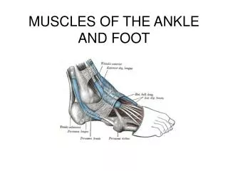
FEEL • TA, heel (Haglaund’s, plantar fasciitis) • Peroneal tendons, lateral ligament, 5th MT base, Tailor’s bunion • Forefoot: Morton’s, MTPJ synovitis, 1st MTPJ, Freiberg’s, stress fractures, sesamoiditis • Midfoot: Kohler’s, acc. Navicular, OA • Ankle: OA, OCD, Tib. Post Tendon, tarsal tunnel • Temperature and pulses • Neurologic: sensation, motor, reflexes
MOVE • Ankle • Subtalar joint • Midfoot • Hallux • Toes • Specific Tendons
COMMON FOOT DISORDERS • Hallux Valgus and Rigidus • Lesser toe deformities • Hammer toe • Mallet toe • Claw toe • Flat foot • Metatarsalgia
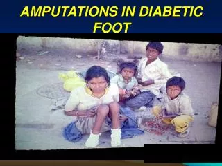
GENERAL PROFILE OF DEFORMITIES • Commonly seen in females • 82% of women report having foot pain, while 72% report one or more foot deformities. • More than 7 out of 10 women develop a bunion, hammertoe, or other painful foot deformity. • Nine out of 10 women’s foot deformities can be attributed to tight shoes.
HALLUX VALGUS Definition Lateral deviation of great toe Aetiology • Familial • Inappropriate footwear • Flatfeet • Long first ray • Incongruous 1st MTP joint articular surface • Metatarsus primus varus • Rheumatoid arthritis. HV HV + Claw toes
HALLUX VALGUS CONTD. Pathogenesis • Complex deformity with angle between 1st & 2nd MT > 9 degrees and valgus angle at MTP joint >20 degrees. • Valgus posture of great toe causing hammer toe like deformity of second toe. • Splaying of forefoot causing bunion. • Incongruence causing osteoarthritis of 1st MTP joint.
WHEN TO REFER Symptoms • Bunion pain • Transfer metatarsalgia • Significant deformity causing: • 2nd toe deformity • Shoe wear problems • Cosmesis – relative contraindication
SIGNS • Bunion and inflamed overlying bursa and skin • Valgus and pronation deformity of hallux. • Painful callus on 2nd toe • Second toe is forced into hyperextension by deviated great toe • Transfer metatarsalgia/thickened skin over MT heads. • Increased valgus angle at first MTP joint • Valgus angle at first MTP joint >20 degrees • Angle between 1st & 2nd MT >9 degrees
MANAGEMENT • Entire foot must be assessed first. • X-ray of foot –Standing dorso plantar, oblique & axial sesamoid views Medial exostosis (bunion) Lateral displacement proximal phalanx Degenerative changes in 1st MTP/IP Joint Intermetatarsal & Hallux Valgus angles
TREATMENT CONSERVATIVE TREATMENT Aim: Relieve pressure over painful bunion prominence • Properly fitted, low heeled stiff-soled shoes • Wide, square shaped toe box • Toe portion stretched to accommodate bunion • Extra-depth shoe to accommodates dorsiflexed second toe • Splint separates first and second toe • Acute pain management • Rest • Apply moist heat • Analgesics
SURGICAL MANAGEMENT Indications • Refractory to conservative management • Severe deformity or bunion pain Factors to be considered before surgery • Valgus deviation of great toe • Varus deviation of first metatarsal • Arthritis of MTP and IP joint • Bunion • Metatarso-cuneiform joint instability • Vascularity & sensibility Surgical Procedures Soft tissue surgery - rarely indicated in adolescent cases Bone/joint procedure remains the gold standard
HALLUX RIGIDUS/DORSAL BUNION Painful limitation of motion at 1st MTP joint Pathogenesis: synovitis, cartilage destruction, osteophyte proliferation, subchondral cysts and sclerosis Clinical presentation: pain, limited dorsiflexion and dorsal osteophyte, dorsal tenderness Aetiology: Trauma, Repeated microtrauma, osteochondritis dissicans and abnormally long first metatarsal
TREATMENT Grade I: Mild osteophytes, joint space preserved • NSAID, orthosis and injection Grade II: Moderate osteophyte formation, joint space narrowing & subchondral sclerosis • Cheilectomy: excision of 20-35% of dorsal metatarsal head aiming for up to 70º of dorsiflexion. Grade III: Severe arthritis • Arthrodesis/joint replacement
LESSER TOE DEFORMITIES • Hammer, Claw and Mallet • Association with HV, RA, DM and NM disorders • Pain, corns, ulcers, shoe wear difficulties • Flexible and fixed • Conservative treatment: manipulation, corn pads, accommodative shoe wear • Surgical treatment: tendon release and transfers for flexible deformities; fusion and excision arthroplasties for fixed deformities.
FLAT FOOT/PES PLANUS Flexible (99%) or Rigid (1%) • Distinguished by Jack test and tiptoeing Pathology - Loss of normal medial longitudinal arch in combination with valgus posture of heel, mild subluxation of subtalar joint & eversion of calcaneum Arch develops till the age of 7-10 years so there is no treatment required 15-20% of adults have asymptomatic pes planus
TREATMENT OF FLEXIBLE PES PLANUS 3-9 years: symptomatic - arch support 10-14 years require investigation Symptomatic patient - rule out accessory navicular or incomplete tarsal coalition and treat accordingly. Adults with painful pes planus not responding to conservative management will benefit with surgery
RIGID PES PLANUS • Aetiology: Congenital vertical talus & tarsal coalition • Tarsal coalition: calcaneo-navicular & talocalcaneal; can be bony, cartilagenous or fibrous. • Symptoms: Foot pain, difficulty walking on uneven surfaces, foot fatigue, peroneal spasm. • Treatment: 4-6 weeks of cast immobilization; surgical treatment includes resection of connecting bar & soft tissue interposition, subtalar arthrodesis, triple arthrodesis.
METATARSALGIA • Pressure from subluxed MTPJs with painful callosities • Freiberg’s AVN (treatment: conservative and surgical) • Stress Fractures • Transfer from first metatarsal insufficiency/HV • Sesamoiditis • Morton’s
MORTON’S METATARSALGIA • Commoner in middle-aged women; 85% unilateral • Aetiology: trauma, ischaemia, entrapment • Pathology: degenerative rather than a true neuroma with perineural fibrosis and demyelination. • Diagnosis: symptom of shooting/constant pain on walking, relieved by rest and removal of footwear; clinical sign of third/second cleft tenderness and palpable click on metatarsal squeeze test. • Treatment: orthoses, injection and excision
ANKLE DISORDERS • Instability • Impingement • Osteochondritis Dissecans of talus • Arthritis • Posttraumatic • Inflammatory • Degenerative
ANKLE INSTABILITY • Repeated acute inversion injuries/laxity • Presentation with pain and instability • Diagnosis: tenderness, anterior draw • Imaging: stress X-rays, MRI • Treatment • Conservative - physiotherapy, splints • Surgical – primary repair/reconstructive procedures
ANKLE IMPINGEMENT • Repeated sporting dorsiflexion injuries • Presentation with anterior ankle pain • Diagnosis: clinical anterior tenderness and ± anterior osteophytes on X-rays • Treatment • Conservative: activity modification/NSAIDs • Surgical: open/arthroscopic decompression
OSTEOCHONDRITIS DISSECANS • Posttraumatic in young patients • Presentation with persistent pain and swelling with stiffness • Diagnosis: clinical tenderness, diffuse swelling • Imaging: X-rays and MRI scan • Treatment: undisplaced lesions treated with rest and cast immobilisation; displaced lesions require arthroscopic removal/drilling
ANKLE ARTHRITIS • Posttraumatic: rare in commonly injured joint; associated with displaced intra-articular fractures and significant lateral ligament complex injury. • Inflammatory: RA in low-demand patients • Degenerative: relatively uncommon • Presentation with pain, swelling, stiffness, limited mobility, limping.
ANKLE ARTHRITIS (CONTD.) • Diagnosis: clinical swelling, tenderness, ↓ROM • Imaging: X-rays, bone scan to assess surrounding joints • Treatment • Conservative: NSAIDs, walking stick, weight reduction and activity modification. • Surgical: arthroscopic/open decompression; ankle arthrodesis (up to 25% non-union, 3 month casting); ankle replacement gives satisfactory mid-term results in properly selected low-demand patients (long-term results?)
TENDON DISORDERS Commonly affected tendons: • Tibialis posterior • Tibialis anterior • Peroneus tendons • Tendoachillis
TIBIALIS POSTERIOR TENDON • Anatomy - posteromedial tendon, origin from posterior surface of tibia & inserts on to the medial cuneiform • Function - plantar flexion, inversion, stabilizes medial longitudinal arch • Important tendon in foot, affection of which causes more functional disability than TA rupture • Aetiology - trauma, chronic flat foot, inflammatory arthropathy, degenerative tendonopathy, chronic tenosynovitis, abnormal insertion, steroid use. • Deformity - collapse of medial longitudinal arch, hindfoot valgus, midfoot abduction, forefoot pronation
PRESENTATION Fatigue of foot with limited activity, medial and lateral pain Flat foot on weight bearing Standing tip toe – heel will go into valgus Clinical examination confirms tenderness, weak/ruptured tendon, hindfoot valgus (flexible/fixed) and a lack of heel varus on tiptoeing Pathogenesis: tenosynovitis, incomplete tear, complete disruption Two groups of patients: Younger patients with inflammatory arthropathy/traumatic rupture Older, typically female patients with degenerative tears PATHOLOGY/PRESENTATION
MANAGEMENT Imaging: X-ray (degeneration), MRI • Tenosynovitis - rest, NSAIDs, short leg walking cast, orthoses, steroid injection in tendon sheath, synovectomy. • Incomplete tear - repair or augmentation with either FDL or FHL. • Complete disruption – repair in traumatic young cases; tendon transfer with medial calcaneal displacement osteotomy (mobile hindfoot) and subtalar/triple arthrodesis (fixed hindfoot). • Satisfactory results in spite of prolonged rehabilitation
TIBIALIS ANTERIOR • Anatomy: • Origin - lateral condyle of tibia, proximal 2/3 of lateral surface of tibia, interosseous membrane • Insertion - base of first metatarsal and medial plantar surface of 1st cuneiform • Action - dorsiflexes and inverts foot • Disorders are common in athletes and old age group • Diagnosis- weakness of dorsiflexion of foot, pain, use of toe extensors for dorsiflexion of foot. • Treatment- steroid injection or synovectomy . Tendon repair rarely required as deformity is not functionally significant.
PERONEAL TENDONS Anatomy: Peroneus longus & brevis are posteolateral tendons originating from fibula and interosseous membrane and are inserted at base of I & V MT respectively.
Pathology: Tenosynovitis- common in high arch foot because of increase in excursion. Sprain/ subluxation - inversion ankle injuries. Symptoms: pain in the outer part of the ankle or just behind the lateral malleolus. This pain commonly worsens with activity and eases with rest. Diagnosis: Examination - tenderness/subluxation X-rays to exclude fracture MRI PERONEAL TENDONS (CONTD.)
Treatment Non-surgical Rest, short-leg walking cast/brace, lateral heel wedge, physical therapy, NSAIDs and Cortisone injection Surgical Tenosynovectomy and repair of split Stabilisation of dislocating tendons by groove deepening, peroneal retinaculum reconstruction and bone block procedures PERONEAL TENDONS (CONTD.)
ACHILLES TENDINITIS/TENDINOSIS • Tendinosis - there will be clinical inflammation, but objective pathologic evidence for cellular inflammation is lacking • Tendinitis - there will be a peritendinous inflammation • Seen in adults in their 30s and 40s • Most commonly affects runners • Heel cord contracture can exacerbate symptoms • Two types: • Non-insertional • Occurs proximal to retrocalcaneal bursa • Generally responds well to non-operative treatment • Insertional • Tenderness is localized to calcaneal tendon insertion • More difficult to treat
Conservative Rest, ice, NSAIDs, physical therapy, orthoses Operative Achilles tendon decompression and debridement if unrelieved by 6 months of conservative measures 90% will have significant relief of symptoms; 10% will have some symptom improvement Complete symptomatic cure not guaranteed TREATMENT
ACHILLES TENDON RUPTURE • Common sporting incidence affecting the young to middle-aged • Mechanism usually involves loading on a dorsiflexed ankle with the knee extended (soleus and gastroc on maximal stretch) or repeated microtrauma • Consider systemic conditions such as gout or hyperparathyroidism (esp. with pure avulsion injury); previous steroid injections • Disabling condition requires approx. 6 months to recover when treated adequately
Diagnosis (suspect in all ankle injury cases): Characteristic history Classical signs: Local tenderness and gap Hyper-dorsiflexion at ankle Thompson/Simmonds test Imaging: Ultrasound and MRI scan in doubtful cases ACHILLES TENDON RUPTURE (CONTD.)
TREATMENT • Non-operative treatment is indicated in older patients and minimally displaced ruptures and involves serial casting over 10-12 weeks (complete equinus, mid equinus, neutral walking). • Operative repair is indicated in younger patients with clinically displaced ruptures, delayed presentation (48-72 hours) and neglected ruptures followed by similar casting regime. • Complications: wound healing and sural nerve injury • Consider DVT prophylaxis
HEEL PAIN • Commonly caused by plantar fasciitis. • Heel spurs often associated. • Pain is worst on waking up. • Causes - obesity, excessive walking/sporting activity, tightplantar fascia & flattening of the arch. • Treatment – orthoses, physical therapy, injection, NSAIDs and (rarely) surgical release in resistant cases.
RETROCALCANEAL BURSITIS • Two bursae: retrocalcaneal (subtendinous) bursa & subcutaneous calcaneal bursa • Causes • Repetitive trauma from shoe wear and sports • Gout, RA and ankylosing spondyloarthropathies • Bursal impingement between the Achilles tendon and an excessively prominent posterior-superior aspect of the calcaneus (Haglund deformity). • Symptoms: pain, swelling, shoe wear difficulty • Signs: tenderness, lump, inflammation
MANAGEMENT • Imaging: X-rays for calcification and Haglund deformity. • Conservative: physical therapy, appropriate shoe wear, injection (risk of tendon rupture). • Surgical Intervention includes resection of Haglund deformity (removal of the calcaneal superoposterior prominence), excision of the painful bursa and debridement of tendon insertion