Download
1 / 21
210 likes | 350 Views
P100 Exams 4224, SV, 2x2x2cm MV Thomas Chong. DW-MRI and MRS to Differentiate Radiation Necrosis and Recurrent Disease in Gliomas. Scans Conducted to Observe MRS Voxel Size Influence on S/N. Single Voxel (SV) Comparisons 1x1x1 (1cm 3 ) 3x3x1 (9cm 3 ) 2x2x2 (8cm 3 ) 3x3x3 (27cm 3 )

E N D
P100 Exams 4224, SV, 2x2x2cm MV Thomas Chong DW-MRI and MRS to Differentiate Radiation Necrosis and Recurrent Disease in Gliomas
Scans Conducted to Observe MRS Voxel Size Influence on S/N • Single Voxel (SV) Comparisons • 1x1x1 (1cm3) • 3x3x1 (9cm3) • 2x2x2 (8cm3) • 3x3x3 (27cm3) • Larger Multi-Voxel (MV) Grid • 2x2x2cm voxels • 6x6 grid
Single Voxel MRS 1x1x1cm Could not get results with metabolite peaks using protocol settings on SV scan. 2-3 attempts • This and subsequent SV positions selected to be in physical middle of hemisphere, away from voids and boundaries. • No discernible metabolite peaks. Why? Seems inconsistent with MV results.
3x3x1cm (9cm3) Single Voxel, S36.6 Exam 4135
3x3x1cm (9cm3) Versus 2x2x2cm (8cm3) Single Voxel, ~S30 Exam 4135 Exam 4139
2x2x2cm (8cm3) Versus 3x3x3cm (27cm3) Single Voxel 2x2x2cm SV, Exam 4139 3x3x3cm SV, Exam 4224
Multivoxel Scan with 2x2x2cm Voxels, Exam 4224, S36.6, Vox30 Note: Spectra shows less background noise that 2x2x2cm SV spectra. Why?
Multivoxel Scan with 2x2x2cm Voxels, Exam 4224, S36.6, Vox 10
Multivoxel Scan with 2x2x2cm Voxels, Exam 4224, S36.6, Vox 12 & 27
INTREPRET Study Protocol • INTERPRET group MRS data protocol • 1.5T GE, Phillips, and Siemens scanners • Both short and long TE • SV volume 4-8cm3; equivalent to cubes of widths 1.6 – 2cm • “whole study protocol took less than 30min” including MRI set for voxel placement, therefore number of scan averages were higher than in our protocol (2). Averaging helps reduce noise. • “N averages metabolites = 192-128” ? • “N averages water = 8-32” ? • Tate A, et al 2006
INTREPRET Study Protocol • Voxel Placement: “Whenever possible voxels were placed entirely within the lesion... avoiding contamination from normal tissue and oedema.” • Data Processing • Some SV spectra created by combining MV voxels. • Custom program based on MRUI software package • 1) Lineshape correction and zero-order phasing using water reference with Klose method • 2) 0.8Hz exponential line-broadening • 3) FFT processing • 4) Water removal by HLSVD; five components removed within +-0.37ppm of water resonance
INTREPRET Study Protocol • Data Processing, cont'd • 4) Water removal by HLSVD; five components removed within +-0.37ppm of water resonance • 5) Residual water suppression; points at 4.2-5.1ppm set to zero • 6) Linear interpolation to 512 points over 1000Hz of Siemens and Phillips data • 7) Spectrum alignment; maximum of choline peak shifted to 3.21ppm • 8) Normalization of spectrum to Euclidian norm of peak heights.
Data-collection Difference Between INTREPRET and Our Protocol • Highest impact difference between protocols appears to be that their scan focused only on region of lesion • INTERPRET used single voxel scans, more averages, larger voxel sizes • These contribute to improve S/N (except maybe the SV vs MV)
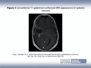
INTERPRET considered mainly glioblastomas, meningiomas, metastases, and astrocytomas grade II
Re-consideration of Current Scan Protocol • Fact: Based on observations of S/N trends with voxel size and theory, we can say that S/N is improved by: • Larger voxel sizes, more averages • Fact: Current protocol gives mostly unusable, low S/N data. We must change it to get data. • Suggestion: • Increase voxel size, increase scan averaging • Reallocate scan time to focus on region of interest; can reduce grid size, # of slices; consider SV
Questions to Discuss When Considering Updating Protocol • If switch to SV's, brains with multiple lesions would require multiple SV scans • If switch to SV's, would be useful to collect data for a non-lesioned “control” voxel at consistent location, e.g. contralateral to lesion region. • Switching to SV's would lose spatially changing metabolite ratio information • Comprise with 3x3 MV grid centered at single lesion? E.g. 1.8x1.8x1.8cm, 3 slices, max avgs to fill protocol scan time. Have to consider scan time tradeoffs. • Must answer why the 1x1x1cm SV test scan did not show any peaks
References • Tate, et al, Development of a decision support system for diagnosis and grading of brain tumours using in vivo magnetic resonance single voxel spectra, NMR in Biomedicine, 2006: 19: 411-434. • Tate, et al, Classification of brain tumours using short echo time 1H MR spectra, J of Magn Reson, 2004: 17: 164-175.