Download

1 / 42
470 likes | 859 Views
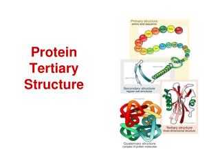
Protein Tertiary Structure Prediction. Structural Bioinformatics. The Different levels of Protein Structure. Primary: amino acid linear sequence. Secondary: -helices, β -sheets and loops. Tertiary : the 3D shape of the fully folded polypeptide chain. PDB: Protein Data Bank.

E N D
Protein Tertiary Structure Prediction Structural Bioinformatics
The Different levels of Protein Structure Primary: amino acid linear sequence. Secondary: -helices, β-sheets and loops. Tertiary: the 3D shape of the fully folded polypeptide chain
PDB: Protein Data Bank • DataBase of molecular structures : Protein, Nucleic Acids (DNA and RNA), • Structures solved by X-ray crystallography NMR Electron microscopy
RCSB PDB – Protein Data Bank http://www.rcsb.org/pdb/
How can we view the protein structure ? • Download the coordinates of the structure from the PDB http://www.rcsb.org/pdb/ • Launch a 3D viewer program For example we will use the program Pymol The program can be downloaded freely from the Pymol homepage http://pymol.sourceforge.net/ • Upload the coordinates to the viewer
Pymol example • Launch Pymol • Open file “1aqb” (PDB coordinate file) • Display sequence • Hide everything • Show main chain / hide main chain • Show cartoon • Color by ss • Color red • Color green, resi 1:40 Help http://pymol.sourceforge.net/newman/user/toc.html
Predicting 3D Structure Outstanding difficult problem Based on sequence homology • Comparative modeling (homology) Based on structural homology • Fold recognition (threading)
Based on Sequence homology Comparative Modeling Similar sequences suggests similar structure
Sequence and Structure alignments of two Retinol Binding Protein
Structure Alignments There are many different algorithms for structural Alignment. The outputs of a structural alignment are a superposition of the atomic coordinates and a minimal Root Mean Square Distance (RMSD) between the structures. The RMSD of two aligned structures indicates their divergence from one another. Low values of RMSD mean similar structures
Dali (Distance mAtrix aLIgnment) DALI offers pairwise alignments of protein structures. The algorithm uses the three-dimensional coordinates of each protein to calculate distance matrices comparing residues. See Holm L and Sander C (1993) J. Mol. Biol. 233:123-138. SALIGN http://salilab.org/DBALI/?page=tools
Based on Sequence homology Comparative Modeling Similar sequence suggests similar structure Builds a protein structure model based on its alignment to one or more related protein structures in the database
Based on Sequence homology Comparative Modeling • Accuracy of the comparative model is related to the sequence identity on which it is based >50% sequence identity = high accuracy 30%-50% sequence identity= 90% modeled <30% sequence identity =low accuracy (many errors)
Homology Threshold for Different Alignment Lengths Homology Threshold(t) Alignment length (L) A sequence alignment between two proteins is considered to imply structural homology if the sequence identity is equal to or above the homology threshold t in a sequence region of a given length L. The threshold values t(L) are derived from PDB
Comparative Modeling • Similarity particularly high in core • Alpha helices and beta sheets preserved • Even near-identical sequences vary in loops
Based on Sequence homology Comparative Modeling Methods MODELLER (Sali –Rockefeller/UCSF) SCWRL (Dunbrack- UCSF ) SWISS-MODEL http://swissmodel.expasy.org//SWISS-MODEL.html
Based on Sequence homology Comparative Modeling Modeling of a sequence based on known structures Consist of four major steps : • Finding a known structure(s) related to the sequence to be modeled (template), using sequence comparison methods such as PSI-BLAST 2. Aligning sequence with the templates 3. Building a model 4. Assessing the model
Based on Structure homology Fold Recognition
Based on Secondary Structure Protein Folds: sequential and spatial arrangement of secondary structures Hemoglobin TIM
Similar folds usually mean similar function Transcription factors Homeodomain
The same fold can have multiple functions Rossmann 12 functions 31 functions TIM barrel
Based on Structure homology Fold Recognition • Methods of protein fold recognition attempt to detect similarities between protein 3D structure that have no significant sequence similarity. • Search for folds that are compatible with a particular sequence. • "the turn the protein folding problem on it's head” rather than predicting how a sequence will fold, they predict how well a fold will fit a sequence
Based on Structure homology Basic steps in Fold Recognition : Compare sequence against a Library of all known Protein Folds (finite number) Query sequence MTYGFRIPLNCERWGHKLSTVILKRP... Goal: find to what folding template the sequence fits best There are different ways toevaluate sequence-structure fit
Potential fold Based on Secondary Structure homology There are different ways toevaluate sequence-structure fit 1) ... 56) ... n) ... ... -10 ... -123 ... 20.5 MAHFPGFGQSLLFGYPVYVFGD...
Based on Secondary Structure homology Programs for fold recognition • TOPITS (Rost 1995) • GenTHREADER (Jones 1999) • SAMT02 (UCSC HMM) • 3D-PSSMhttp://www.sbg.bio.ic.ac.uk/~3dpssm/
Ab Initio Modeling • Compute molecular structure from laws of physics and chemistry alone Theoretically Ideal solution Practically nearly impossible WHY ? • Exceptionally complex calculations • Biophysics understanding incomplete
Ab Initio Methods • Rosetta (Bakers lab, Seattle) • Undertaker (Karplus, UCSC)
CASP - Critical Assessment of Structure Prediction • Competition among different groups for resolving the 3D structure of proteins that are about to be solved experimentally. • Current state - • ab-initio - the worst, but greatly improved in the last years. • Modeling - performs very well when homologous sequences with known structures exist. • Fold recognition - performs well.
What can you do?FOLDITSolve Puzzles for Science A computer game to fold proteins http://fold.it/portal/puzzles
What’s Next Predicting function from structure
Structural Genomics: a large scale structure determination project designed to cover all representative protein structures ATP binding domain of protein MJ0577 Zarembinski, et al., Proc.Nat.Acad.Sci.USA, 99:15189 (1998)
Wanted ! Automated methodsto predict function from the protein structures resulting from the structural genomic project. As a result of the Structure Genomic initiative many structures of proteins with unknown function will be solved
Approaches for predicting function from structure ConSurf - Mapping the evolution conservation on the protein structure http://consurf.tau.ac.il/
Approaches for predicting function from structure PFPlus – Identifying positive electrostatic patches on the protein structure http://pfp.technion.ac.il/
Approaches for predicting function from structure SHARP2 – Identifying positive electrostatic patches on the protein structure http://www.bioinformatics.sussex.ac.uk/SHARP2
Machine learning approach for predicting function from structure Find the common properties of a protein family (or any group of proteins of interest) which are unique to the group and different from all the other proteins. Generate a model for the group and predict new members of the family which have similar properties.
Knowledge Based Approach Basic Steps 1. Building a Model • Generate a dataset of proteins with a common function (DNA binding protein) • Generate a control dataset • Calculate the different properties which are characteristic of the protein family you are interested for all the proteins in the data (DNA binding proteins and the non-DNA binding proteins • Represent each protein in a set by a vector of calculated features and build a statistical model to split the groups
Basic Steps 2. Predicting the function of a new protein • Calculate the properties for a new protein And represent them in a vector • Predict whether the tested protein belongs to the family
TEST CASE Y14 – A protein sequence translated from an ORF (Open Reading Frame) Obtained from the Drosophila complete Genome >Y14 PQRSVGWILFVTSIHEEAQEDEIQEKFCDYGEIKNIHLNLDRRTGFSKGYALVEYETHKQALAAKEALNGAEIMGQTIQVDWCFVKG G
? Support Vector Machine (SVM) To find a hyperplane that maximally separates the RNA-binding from non-RNA binding into two classes RNA binding =[x1, x2, x3…] Kernel function new protein structure Non-NA binding =[y1, y2,y3…] Input space Feature space
>Y14 PQRSVGWILFVTSIHEEAQEDEIQEKFCDYGEIKNIHLNLDRRTGFSKGYALVEYETHKQALAAKEALNGAEIMGQTIQVDWCFVKG G Y14 DOES NOT BIND RNA