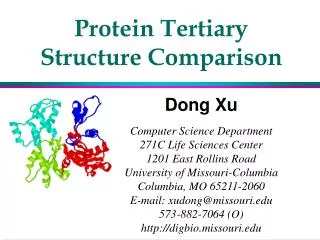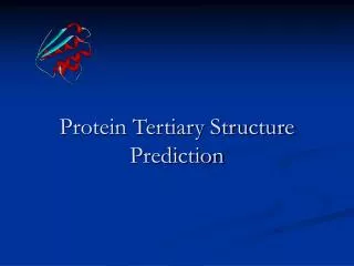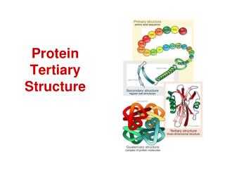
Protein Tertiary Structure
Protein Tertiary Structure. What to Know. What are some protein functions? General principles for protein folding General structural features of globular and structural proteins Know the 4 common globular protein motifs

Protein Tertiary Structure
E N D
Presentation Transcript
What to Know • What are some protein functions? • General principles for protein folding • General structural features of globular and structural proteins • Know the 4 common globular protein motifs • Understand how protein structures are stabilized and why some portions of proteins are marginally stable • Why is protein flexibility and motion important?
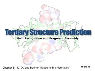
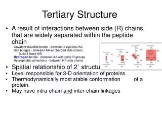
TERTIARY STRUCTURE OF PROTEINS • THE THREE-DIMENSIONAL CONFORMATION OF A PROTEIN • The overall spatial relationships between segments of primary and secondary structure • The scope of tertiary structure is thus long-range • Determined by x-ray diffraction and NMR
Globular Protein Functions • Functionally this group is most diverse: • Enzymes • Transport proteins • Antibodies • Cell surface proteins • Integral membrane proteins • Virus coat proteins • Storage proteins • DNA binding proteins • ????
How Do Polypeptides Fold into Three-Dimensional Protein Structures? Several important principles: • Secondary structures form wherever possible (due to formation of large numbers of H bonds) • Helices and sheets often pack close together • Peptide segments between secondary structures tend to be short and direct • Proteins fold so as to form the most stable structures. Stability arises from: • Formation of large numbers of intramolecular hydrogen bonds • Reduction in the surface area accessible to solvent that occurs upon folding
How Do Polypeptides Fold into Three-Dimensional Protein Structures? • Two factors lie at the heart of these principles: • Proteins are typically a mixture of hydrophilic and hydrophobic amino acids • The hydrophobic groups tend to cluster together in the folded interior of the protein
Tertiary Structure Several important principles: Proteins fold to make the most stable structures. Globular proteins. Create maximum internal bonds and minimize solvent contact. Fibrous proteins. Create maximum intermolecular bonds and maximize molecule to molecule contact.
FIBROUS PROTEINS Characterized by association of helical chains which coil around each other forming coiled-coils. Keratin and Myosin (heptad repeat) Collagen (triplet repeat, G-X-Y, G-P-P)
Structure of Collagen Triple helical structure: ●Three αchains each coiled in left-handed sense (Minor helix). ●Three α Chains coiled about each other in right-handed sense (Major helix).
H-bonds in the collagen foldFrom Gly-N groups to C=O function of the preceding amino acid in a neighboring chain. Also, hydroxyproline participates.
Globular Proteins • Most polar residues face the outside of the protein and interact with solvent • Most hydrophobic residues face the interior of the protein and interact with each other • Packing of residues is close • However, ratio of VdW volume to total volume is only 0.72 to 0.77, so empty space exists • The empty space is in the form of small cavities"Random coil" is not random • Structures of globular proteins are not static • Various elements and domains of protein move to different degrees • Some segments of proteins are very flexible and disordered
Most Globular Proteins Belong to One of Four Structural Classes • Proteins can be classified according to the type and arrangement of secondary structure • There are four classes: • All α proteins, in which α helices predominate • All β proteins, in which β sheets predominate • α/β proteins, in which helices and sheets are intermingled • α+β proteins, which contain separate α-helical and β-sheet domains
The Serine Proteases Trypsin, chymotrypsin, elastase, thrombin, subtilisin, plasmin, TPA • All involve a serine in catalysis - thus the name • Ser is part of a "catalytic triad" of Ser, His, Asp • Serine proteases are homologous, but locations of the three crucial residues differ somewhat • Enzymologists agree, however, to number them always as His-57, Asp-102, Ser-195
Comparison of the amino acid sequences of chymotrypsinogen, trypsinogen, and elastase. Filled circles indicate residues that are identical in all three proteins. The positions of the three catalytically important active-site residues (His57, Asp102, and Ser195) are indicated.
Structure of chymotrypsin (white) in a complex with eglin C (blue ribbon structure), a target protein. The residues of the catalytic triad (His57, Asp102, and Ser195) are highlighted. His57 (blue) is flanked above by Asp102 (red) and on the right by Ser195 (yellow). Catalytic site is filled by a peptide segment of eglin. Note how close Ser195 is to the peptide that would be cleaved in a chymotrypsin reaction.
Parvalbumin Bob Kretsinger
Globular Proteins The structure of ribonuclease, showing elements of helix, sheet and random coil.
Protein surfaces are complex The surfaces of proteins are complementary to the molecules they bind.
Waters on the Protein Surface Stabilize the Structure The surfaces of proteins are ideally suited to form multiple H bonds with water molecules.
Coiled Coils: More stable than single helix or two straight rod-like helices Francis Crick
Four helix bundle is common in domain structure proteins Myohemerythrin, cytichromec’,ferritin, coat protein of TMV
Beta-alpha-beta proteins: triosphosphate isomerase and co-enzyme binding domain of a dehydrogenase: beta stands are parallel
Methylmalonyl-coenzyme A mutase Inside of the barrel is lined with hydrophillic side chains (S and T) which creates a hole for substrate to bind
Examples of Open twisted α/β structures: Flavodoxin and adenylate kinase
Denaturation Leads to Loss of Protein Structure and Function • The cellular environment is suited to maintaining the weak forces that preserve protein structure and function • External stresses – heat, chemical treatment, etc. – can disrupt these forces in a process termed denaturation – the loss of structure and function • The cooking of an egg is an everyday example • Ovalbumin, the principal protein in egg white, remains in its native structure up to a characteristic melting temperature, Tm determined via differential scanning calorimetry (DSC) • Above this temperature, the structure unfolds and function is lost