Fig. 15.4
180 likes | 360 Views
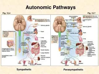
Autonomic Pathways. Fig. 15.4. Fig. 15.7. Sympathetic. Parasympathetic. Sympathetic thoracolumbar fight or flight mass activation possible e.g., vasoconstriction and heart rate blood pressure blood flow to skin and viscera blood flow to skeletal muscles

Fig. 15.4
E N D
Presentation Transcript
Autonomic Pathways Fig. 15.4 Fig. 15.7 Sympathetic Parasympathetic
Sympathetic thoracolumbar fight or flight mass activation possible e.g., vasoconstriction and heart rate blood pressure blood flow to skin and viscera blood flow to skeletal muscles e.g., [glucose]blood due to glycogenolysis Parasympathetic craniosacral “vegetative” functions no mass activation individual responses e.g., heart rate e.g., GI activity secretion motility Two Divisions of the ANS
Sympathetic and Parasympathetic • antagonistic effects • e.g., heart rate • no rule about which stimulates and which inhibits • separate effects • e.g., sympathetic: myocardial contractility parasympathetic: no effect on contractility • cooperative effects • e.g., sexual function
Sympathetic Pathways • Preganglionic fibers • leave spinal cord via spinal nerves T1 – L2 • leave spinal nerve, travel to a ganglion of the sympathetic trunk (chain) and split into branches • various branches synapse at that ganglion ascend or descend to another trunk ganglion and synapse there pass through the trunk to a prevertebral ganglion and synapse • celiac, superior mesenteric and inferior mesenteric ganglia • Different body segments can be simultaneously stimulated. • Mass activation is possible. Fig. 15.5
Sympathetic Pathways Fig. 15.4 • Postganglionic fibers • return to nerves • travel in nerves to target organs • Note: The adrenal medulla is like a sympathetic ganglion. Adrenal medullary cells are like postganglionic neurons. to blood: epinephrine norepinephrine
Parasympathetic Pathways Fig. 15.7 • Long preganglionic fibers travel in cranial nerves and in splanchnic nerves from the sacral region to parasympathetic ganglia in or near organs. • vagus nerve (X) • heart, lung, upper GI • pelvic splanchnic nerves • defecation, urination, sexual responses • Short postganglionic fibers spread out into organ.
Neurotransmitters • acetylcholine and norepinephrine • Rules: • Neurons are named by the main neurotransmitter they secrete. • adrenergic fibers • secrete norepinephrine (noradrenaline) • cholinergic fibers • secrete acetylcholine • Membrane receptors are named by the neurotransmitter they receive. • adrenergic receptors • receive norepinephrine and epinephrine (adrenaline) • cholinergic receptors • receive acetylcholine
Cholinergic and Adrenergic Fibers Sympathetic Usually NE ACh Target cell Preganglionic neuron ACh Occasionally: e.g., to sweat glands Postganglionic neuron Parasympathetic ACh Target cell ACh Postganglionic neuron Preganglionic neuron Fig. 15.8
Fiber Types • Simplistic associations • postganglionic neurons • sympathetic: adrenergic • parasympathetic: cholinergic • Detailed rules • adrenergic fibers • most sympathetic postganglionic neurons • norepinephrine • adrenal medullary cells • epinephrine (80%) • norepinephrine (20%)
Fiber Types • Detailed rules (cont’d) • cholinergic fibers • ALL preganglionic neurons • both sympathetic and parasympathetic • parasympathetic postganglionic neurons • a few postganglionic sympathetic neurons • to eccrine sweat glands • to blood vessels supplying skeletal muscle
Receptor Types Sympathetic Usually Adrenergic receptors: 1, 1, 2 Nicotinic cholinergic receptors NE ACh Target cell Preganglionic neuron ACh Muscarinic cholinergic receptors Postganglionic neuron Occasionally: e.g., to sweat glands Nicotinic cholinergic receptors Parasympathetic ACh Target cell ACh Muscarinic cholinergic receptors Postganglionic neuron Preganglionic neuron Fig. 15.8
Receptor Types • Cholinergic Receptors • nicotinic cholinergic receptors • channel-linked receptor / ligand-gated channel • location • cell bodies and dendrites of all postganglionic neurons • [neuromuscular junction of skeletal muscle fibers] • muscarinic cholinergic receptors • G-protein linked • work via intracellular (“second”) messengers • location • parasympathetic effector tissue
Receptor Types • Adrenergic Receptors • many and subtypes • for this course • 1, 1 and 2 adrenergic receptors • 2 is important for pharmacology, but will not be discussed in A&P. • All are G-protein linked. • location: • sympathetic effector tissue; examples: • 1: most vascular smooth muscle (vasoconstriction) • 1: heart ( heart rate, myocardial contractility) • 2: bronchiolar smooth muscle, vascular smooth muscle of skeletal muscle blood vessels (bronchiolo- and vasodilation)
Autonomic Pharmacology • Various drugs can • act presynaptically to stimulate or inhibit the release of neurotransmitters • and/or • act postsynaptically to be agonists or antagonists to the different receptor types on the receiving cells. • For example, • propranolol: -blocker • phenylephrine: -agonist Fig. 15.8
Regulation of ANS Activity • The ANS always has some activity (“tone”). • There is always some release of neurotransmitter. • Regulation is not a matter of turning the neurons on or off, but of increasing or decreasing their secretion. • sympathetic tone / sympathetic tone • parasympathetic tone / parasympathetic tone
Regulation of ANS Activity • Reflexes • via spinal cord and brainstem • e.g., cardiovascular, respiratory, micturition, defecation and sexual reflexes • Higher Brain Influences • via hypothalamus, limbic system, cerebral cortex • e.g., to activate “fight or flight”