Download
1 / 22
220 likes | 529 Views
SYNOVIAL ENTRAPMENT SYNDROMES. Concepts & Treatment – Cineradiographic & Arthrographic demonstrations of a new diagnosis.

E N D
SYNOVIAL ENTRAPMENT SYNDROMES Concepts & Treatment – Cineradiographic & Arthrographic demonstrations of a new diagnosis
Synovial entrapment is common- and the cause of many painful conditions involving virtually any diarthrodial joint. In my experience, it is a reason for dramatic relief after a joint gapping type of manipulative procedure.
This dissection by Dr. Wolfgang Rauschning demonstrates normal synovial tags in thoracic vertebral joints, joints of the spine in the area of the ribs. The tags secrete lubricating fluid. • This tissue is laden with nerve fibers, and if they become entrapped, severe pain can result, which cannot be relieved until the tissue is released.
The next slide simulates Pat’s position in bed, wearing the hard cervical collar, as she rested.
Photo of Pat revealing the pain pattern that developed. The intense pain on the top of her shoulder directly over the acromio-clavicular joint is not evident here.
The arthrogram of Pat’s presumed normal left AC joint. The “reservoir” at the inferior aspect of the joint is obvious, as is the vertical column of solution inside the joint, demonstrating the absence of entrapped soft tissue.
Arthrogram of Pat’s symptomatic right side. A reservoir is again present, but notice the paucity of dye in the joint. Clearly, soft tissue is entrapped that is preventing the dye from entering. • (This was performed in 1974 and is possibly the 1st clinical AC arthogram performed.)
This arthrogram on fresh cadaver material, one of fifty from the 1st to 10th decades, demonstrates a “classical” normal AC appearance.
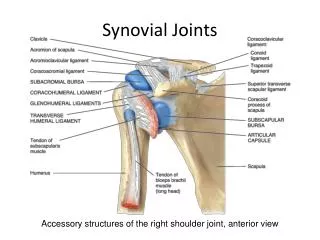
The ligaments of the AC joint • This drawing requires some knowledge to fully appreciate. It demonstrates that all the ligaments restrain the clavicle from moving superiorly. None act to limit its downward movement.
With Pat on her right side, the hard collar pushing down on her clavicle, the force of the bed tending to stretch the deltoid muscle, allowing the humerus to move inferiorly – with a weak, essentially nonexistent inferior joint capsule – the stage was set for sudden inferior dislocation of the clavicle under the acromion.
This view of the side lying skeleton (sorry, it’s reversed) reveals the clavicle as a vertical strut. When it “sprung,” it not only caught synovium and trapped it in the joint as it spontaneously reduced, it’s sudden loss as a support of the thoracic spine caused the spine to “buckle” and become locked in severe dysfunction, possibly with entrapped synovium, as well.
The following cinearthrogram of Lisa’s left hip is essentially self explanatory. The soft tissue is seen entrapped. The motion of the hip “disarticulation” on axial traction was abnormal. The impulsive hip extraction almost certainly was curative. The subsequent arthrogram reveals a normal joint space. The “pressure” aspects of the arthrogram are explained in the text.
Consider soft tissue entrapment syndrome. It is usually easy to diagnose. Manipulative therapy – distracting the joint – usually provides gratifying results. • Be well.