Download
1 / 30
300 likes | 335 Views
Explore weight encoding criteria and methods using internal fluorophore and fluorescence resonance energy transfer in DNA-based perceptron systems. Discover competitive hybridization for signal output proportional to mRNA concentration.

E N D
Weight Encoding Methods in DNA Based Perceptron 2004. 7. 6 임희웅
Contents • Weight Encoding Criteria • Methods • Internal fluorophore • Fluorescence resonance energy transfer • Competitive hybridization of labeled and unlabeled probe • Materials
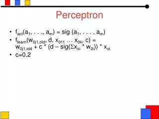
Weight Encoding Criteria • Output • Fluorescence Signal • Proportional to • Concentration of corresponding mRNA (xi) • Corresponding of weight value (ti) Assumption: Final signal (output) is the sum of each output signal Outputi
Internal Fluorophore Absolute Weight Value High Low (+) Fluorophore 1 Sign (-) Fluorophore 2 More fluorophores Less fluorophores
Fluorescence Resonance Energy Transfer • Fluorescence Resonance Energy Transfer • Transfer of the excited state energy from the initially excited donor (D) to an acceptor (A) • Distance-dependent interaction between donor and acceptor without emission of a photon
Quencher • Small weight short probe length weak signalLarge weight long probe length strong signal Absolute Weight Value High Low (+) Fluorophore 1 Sign (-) Fluorophore 2 Longer probe Shorter probe
FRET Efficiency • Förster Distance (R0) • A distance where the efficiency is 50%.
DNA Helix Model for FRET Efficiency Clegg et al. PNAS 1993
Our Dye-Quencher Pairs • How Many? • 2 pairs for (+) and (-) • Condition • Dark quencher • No background signal, high S/N ratio • Discrimination between (+) and (-) • Large difference in wavelength peak • Multiplexing • Small interference between two dyes. • How about using the pairs in RT-PCR? • Sensitivity • Efficient sensitivity in the concentration of experimental condition. • Enough Förster Distance (R0) • Active range of FRET corresponding to probe length • 3.3Å for one bp,
6FAM cy5 Rox (Texas-red) Hex And BHQ series as dark quencher
More to Know • Fluorescence intensity vs. concentration • Multiplexing
Competitive hybridization of labeled and unlabeled probe • Small weight low ratio of labeled probe weak signalLarge weight high ratio of labeled probe strong signal Unlabeled Probe Labeled Probe Excess Probe Competitive hybridization mRNA
Sign Encoding with One Fluorophore probe Positive weight mRNA Negative weight High Conc. Low Conc.
Reference • Clegg et al. PNAS vol. 90, p 2994~2998, 1993 • Observing the helical geometry of double-stranded DNA in solution by fluorescence resonance energy transfer • Dietrich et al. Reviews in Molecular Biotechnology, vol. 82, p 221~231, 2002 • Fluorescence resonance energy transfer (FRET) and competing processes in donor-acceptor substituted DNA strands: a comparative study of ensemble and single-molecule data
Dual Labeled Probe (1) Bioneer http://www.bioneer.co.kr/biomall/mall_oligo.jsp
Dual Labeled Probe (2) 5’/3’ 1. FAM/BHQ-1 Fluorogenic Probe 2. HEX/BHQ-1 Fluorogenic Probe 3. TET/BHQ-1 Fluorogenic Probe 4. MAX/BHQ-1 Fluorogenic Probe 5. Cy5/BHQ-3 Fluorogenic Probe 6. Cy5/BHQ-2 Fluorogenic Probe 7. Cy3/BHQ-2 Fluorogenic Probe 8. TAMRA/BHQ-2 Fluorogenic Probe 9. ROX/BHQ-2 Fluorogenic Probe Synthegen http://www.synthegen.com/
Dual Labeled Probe (3) IDT http://www.idtdna.com/program/catalog/Dual_Labeled_Fluorescent_Probes.asp
Förster Distance (R0) HiLyte Biosciences, Inc.
http://www.biosearchtech.com/ This chart shows the absorption spectra of all three BHQ dyes (conjugated to poly-T 9-mers and normalized to the T9 absorbance at 260 nm) with the emission maximum of many commonly used reporter groups indicated.