Chapter 29 Human Development
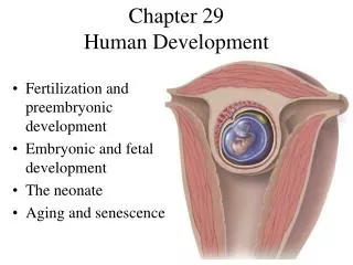
Chapter 29 Human Development. Fertilization and preembryonic development Embryonic and fetal development The neonate Aging and senescence. Sperm Migration. Majority of sperm do not make it to egg destroyed by vaginal acid fail to penetrate the cervical canal mucus

Chapter 29 Human Development
E N D
Presentation Transcript
Chapter 29Human Development • Fertilization and preembryonic development • Embryonic and fetal development • The neonate • Aging and senescence
Sperm Migration • Majority of sperm do not make it to egg • destroyed by vaginal acid • fail to penetrate the cervical canal mucus • go up wrong uterine tube • Move by lashing of sperm tail • Assisted by female physiology • strands of cervical mucus • uterine contractions • chemical attraction
Capacitation • Spermatozoa can reach uterine tube within 10 minutes of ejaculation • can not fertilize egg until undergo capacitation • requires 10 hours for process to occur • female fluids wash away cholesterol & inhibitory factors • sperm membrane becomes fragile & permeable to Ca+2 • Sperm fertile for 48 hours after ejaculation • Conception of a child is optimal if sperm are deposited anytime from 48 hours before ovulation to 14 hours after
Fertilization • Acrosomal reaction of capacitated sperm • release of enzymes from many sperm needed to penetrate granulosa cells and finally the zona pellucida surrounding the egg • hyaluronidase and acrosin • membranes of 2 gametes fuse & sperm enters • Prevention of polyspermy • fast block = depolarization of membrane (by opening of Na+ channels) prevents binding of second sperm • slow block = sperm penetration triggers Ca+2 inflow, causes cortical reaction (secretion from cortical granules forms fertilization membrane)
Fertilization • Secondary oocyte completes meiosis only if fertilized • produces 2nd polar body • Swollen sperm & egg nuclei called pronuclei • Pronuclei rupture • Chromosomes of 2 gametes mix • Fertilized egg is now called a zygote NOTE: location of egg during fertilization
Preembryonic Stage = First 2 Weeks • Cleavage = mitotic divisions that occur for 3 days after fertilization • within 30 hrs – 2 cell stage • zygote splits in half into 2 daughter cells (blastomeres) • within 72 hrs – morula stage (solid ball of small cells) • Morula free in uterine cavity for 4-5 days • nourished by endometrial secretion (uterine milk) • Zona pellucida disintegrates to release blastocyst • outer cells of hollow sphere = trophoblast which helps to form placenta • inner cell mass (embryoblast) develops into embryo
Twins • Dizygotic (fraternal) twins • 2 eggs are ovulated and fertilized (2 zygotes) • as different as any other siblings • Monozygotic (maternal) twins • 1 egg is fertilized (1 zygote) but embryoblast splits into two • genetically identical siblings (must be same sex)
Implantation of Blastocyst • Attaches to uterine wall 6 days after ovulation • Syncytiotrophoblast is the multinucleate mass that grows “roots” and digests its way into endometrium • secretes human chorionic gonadotropin (HCG) • stimulates corpus luteum to continue to secrete hormones to maintain endometrium and prevent menstruation (detectable by urine test kit) • becomes chorion • Endometrium completely encloses implanting embryo
Steps of Embryogenesis • Arrangement of blastomeres into 3 primary germ layers • Formation of amniotic cavity between embryoblast & cytotrophoblast (see next slide) • Flattening of embryoblast into embryonic disc formed from ectodermal & endodermal cells • Cells sink into primitive streak (a groove) & spread laterally as the mesoderm layer • gelatinous tissue (mesenchyme cells)
Ectopic Pregnancy • Blastocyst implants somewhere other than uterus • 1 out of 300 pregnancies • most cases occur in uterine tube (tubal pregnancy) • occurs because of tubal obstruction from previous pelvic inflammations, repeated abortions or tubal surgery • Tube can not expand enough & ruptures by 12 weeks • conceptus may reimplant in abdominopelvic cavity • anywhere it finds an adequate blood supply • usually requires an abortion • 9% of abdominal pregnancies result in live birth by cesarian section
Embryonic Stage or Weeks 2 to 9 • Begins when all 3 primary germ layers are present • Conceptus forms a set of membranes external to the embryo • Embryo begins receiving its nutrients from the placenta • Germ layers differentiate into organs and organ systems • presence of organs marks the beginning of fetal stage
Prenatal Nutrition • Trophoblastic nutrition - conceptus is nourished by digestion of endometrial cells (first 8 weeks) • progesterone stimulates decidual cells of uterus • Placental nutrition - conceptus is nourished from mother’s bloodstream through the placenta
Placentation • Formation of the placenta occurs from 11 days to 12 weeks • Chorionic villi • extensions of syncytiotrophoblast into endometrium by digestion & growth of “roots” of tissue • mesenchyme extends into chorionic villi to form embryonic blood vessels • Placental sinus • pools of maternal blood that merge and surround villi • blood stimulates rapid growth of chorionic villi
The Placenta • Once fully developed is disc of tissue 20 cm in diameter and 3 cm thick • Surface facing fetus is smooth & connected to fetus by umbilical cord • Uterine surface consists of villi and decidua basalis region of endometrium • Fetal & maternal blood do not mix • Placental conductivity increases as villi grow since their membranes become thinner • substances pass through by diffusion, facilitated diffusion, active transport and receptor-mediated endocytosis
Embryonic Membranes • Amnion - transparent sac filled with fluid • protects embryo from trauma, temperature changes, adhesions and provides freedom of movement • forms from maternal plasma filtrate & fetal urine • at term, amnion contains 700 to 1000 mL of fluid • Yolk sac - hangs from ventral side of embryo • contribute to GI tract, blood cells and germ cells • Allantois - foundation of umbilical cord & urinary bladder • Chorion - outermost membrane • chorionic villi form fetal portion of the placenta
Organogenesis • Formation of organs & organ systems from primary germ layers • at 8 weeks, all organs are present in 3 cm long fetus • heart is beating and muscles exhibit contracts • Derivatives of the ectoderm • epidermis, nervous system, lens & cornea, internal ear • Derivatives of the mesoderm • skeleton, muscle, cartilage, blood, lymphoid tissue, gonads & ducts, kidneys & ureters • Derivatives of the endoderm • gut & respiratory epithelium & glands, bladder & urethra
Fetal Development & Circulation • Fetus = from 8 weeks until birth • organs mature to support external life • Anatomical changes in fetal circulation • spaces in mesoderm become lined with endothelium & merge into blood vessels and lymphatic vessels • side-by-side endothelial tubes fuse to form heart • Fetal circulation • umbilical-placental circuit via umbilical cord • circulatory shunts • ductus venosus connects to inferior vena cava • foramen ovale connecting right & left atria • ductus arteriosus connects pulmonary trunk to aorta
The Neonate or Newborn • Transitional period • first 6-8 hours heart & respiratory rate & body temperature falls • periods of sleeping & gagging on mucus & debris • feed every 3 to 4 hours during 6 week neonatal period • Respiratory adaptations of newborn • onset of breathing due to CO2 accumulation • great effort to inflate lungs for first few breaths • Immunological adaptation • maternal antibody, IgG, diffuses across placenta • provides 6 mo of protection from most infectious diseases while fetal production • IgA in breast milk can protect newborn from gastroenteritis
Circulatory Adaptations • Umbilical arteries and veins become ligamentous • Ligamentum venosum (liver) • Fossa ovalis (heart) • Ligamentum arteriosum (vessels)
Thermoregulation & Fluid Balance • Infant has larger ratio of surface area to volume • loses heat more easily • defenses • brown fat deposited during weeks 17 to 20 fetal life • mitochondria breakdown pyruvic acid & release only heat • grows and increases metabolic rate • accumulates subcutaneous fat • Kidneys not fully developed at birth • can not concentrate urine so have a high rate of water loss & require more fluid intake, relative to body weight
Premature Infants • Infants born weighing under 5.5 lb. • Infants born before 7 months suffer from • respiratory distress syndrome • insufficient surfactant causing alveolar collapse with exhalation • thermoregulatory problems due to undeveloped hypothalamus -- keep in incubator • digestive system not well developed must be fed low-fat formula instead of breast milk • immature liver fails to synthesize plasma proteins • edema, deficiency of clotting & jaundice from bile
Congenital Anomalies • Infectious diseases • microorganisms that can cross the placenta include • herpes simplex, rubella, cytomegalovirus, HIV • results range from mild effects to blindness, cerebral palsy & severe physical & mental retardation are just some of the results • Teratogens are viruses, chemicals or other agents that cause anatomical deformities in fetus • thalidomide (unformed arms or legs) • fetal alcohol syndrome, smoking & X rays • cardiac & CNS defects, anencephaly, cleft lip and palate, hyperactivity and poor attention span
Effects of Thalidomide Sleeping medication taken early in pregnancy with severe teratogen effects on limb development.
Mutagens and Genetic Anomalies • Mutagen is any agent that alters DNA or chromosome structure • radiation or diverse chemicals • Most common genetic disorders from failure of homologous chromosomes to separate during meiosis (normal separation = disjunction) • Nondisjunction – unequal # of chromosomes go to daughter cells causing aneuploidy (wrong #) • can be detected prior to birth with amniocentesis (examining fetal cells from amniotic fluid) or chorionic villus sampling (examine placental cells)
Nondisjunction & Aneuploidy • Nondisjunction of sex chromosomes • Triplo-X syndrome (XXX) -- egg receiving 2 X chromosomes fertilized by X carrying sperm • infertile female with mild intellectual impairment • Klinefelter syndrome (XXY) -- egg receiving 2 X chromosomes fertilized by Y carrying sperm • sterile males with average intelligence (undeveloped testes) • Turner syndrome (XO) -- egg receiving no X chromosomes but fertilized by X carrying sperm • sterile, webbed neck, female with no 2nd sexual features • Nondisjunction of autosomes – often lethal • Most survivable type is Down syndrome (trisomy-21)
Down Syndrome Characteristics • Effects of carrying 3 copies of chromosome 21 include: short stature, flat face with epicanthal folds on eyes, enlarged tongue, stubby fingers and mental retardation • Occurs in proportion to age of mother • 1 in 9 for a 48 year old mother; 1 in 3000 for under 30y/o mother
Aging and Senescence • Aging is all changes occurring with the passage of time -- growth, development & degeneration • Senescence is the degeneration that occurs after the age of peak functional efficiency • leading causes of death from 18 to 34 is accidents, homicides, suicides and AIDS • leading causes of death after 55 is senescence related • cancer, stroke, diabetes, heart & lung disease • All organ systems do not degenerate at the same rate - some changes not evident except under stress
Aging of Integumentary System • Becomes noticeable in late 40s • Intrinsic aging of skin • gray, thinning, dry hair • paper-thin, loose skin that sags • skin that bruises easily & heals slowly • hypothermia in cold weather & heat stroke in hot • atrophy of cutaneous vessels, sweat glands & subcutaneous fat • vitamin D production Ca+2 deficiency • Photoaging is degeneration in proportion to UV exposure -- skin spots, skin cancer, wrinkling
Aging of Skeletal System • Osteopenia is loss of bone mass • after 30, osteoblasts less active than osteoclasts • after 40, women loose 8% per decade; men 3% • brittle bones fracture & heal slowly due to protein synthesis • Joint diseases • synovial fluid less abundant & articular cartilage thinner or absent -- friction causes pain • osteoarthritis is common cause of physical disability • breathing difficult due to calcification of sternocostal jts. • but herniated discs less common (less nucleus pulposus)
Aging of Muscular System • Muscular atrophy causes replacement of lean body mass (muscle) with fat • by 80, we have half as much strength & endurance • fast-twitch fibers exhibit earliest & most severe atrophy • Reasons for loss of strength • fibers have fewer myofibrils, smaller mitochondria, less enzymes, glycogen & myoglobin • fewer motor neurons in spinal cord with less efficient synaptic transmission of acetylcholine • sympathetic nervous system is less efficient so less efficient blood flow to muscles causes fatigue
Aging of Nervous System • Cerebral & neuronal atrophy • from age 35 on, 100,000 brain cells die every day • brain weight 50% less by age 75 • cortex thinner, gyri narrower, fewer synapses & neuroglia, less neurotransmitter & receptors • degeneration of myelin slows down signal conduction • neurons contain less ER and Golgi as their metabolism slows • accumulate more lipofuscin pigment, neurofibrillary tangles • extracellular protein plaques accumulate • Motor coordination, intellectual function & short-term memory suffer the most • Autonomic nervous system is less efficient at regulating body temperature & blood pressure
Aging of the Sense Organs • Vision • loss of flexibility of lenses (presbyopia) • cataracts or cloudiness of lenses • night vision is impaired due to fewer receptors, vitreous body less transparent, pupil dilators atrophy & enzymatic reactions become slower • glaucoma risks increase with other structural changes • Hearing • tympanic membrane & ossicle joints stiffen • hair cells & auditory nerve fibers die • death of receptor cells in semicircular duct cells result in poor balance & dizziness • Taste and smell is blunted as receptors decline in number
Aging of Endocrine System • Degenerates less than any other system • only reproductive, growth & thyroid hormones decline steadily after adolescence • other hormones secreted at fairly stable rate • target cell sensitivity may decline • Pituitary gland is less sensitive to negative feedback inhibition by adrenal glucocorticoids • response to stress is prolonged • Type II diabetes is more common • more body fat insulin sensitivity of other cells • target cells have fewer insulin receptors
Aging of Circulatory System • Anemia may result from nutrition, lack of exercise, changes in erythropoiesis, lack of intrinsic factor vitamin B12 absorption • Coronary atherosclerosis leads to angina, infarction, arrhythmia and heart block • heart walls thinner, stroke volume & output declines • degeneration of nodes and conduction system • Atherosclerosis of other vessels increases BP • vessels stiffen & can not expand as effectively • Varicose veins due to weaker valves • pooling of blood raises capillary BP causing edema
Aging of Immune System • Amounts of lymphatic tissue & red bone marrow decline • fewer hemopoietic stem cells, disease-fighting leukocytes & antigen-presenting cells • Lymphocytes fail to mature • Both types of immune responses are less efficient • less protection from cancer & infectious disease
Aging of Respiratory System • Declining pulmonary ventilation • costal cartilages less flexible • lungs have less elastic tissue & fewer alveoli • Elderly less able to clear lungs of irritants & pathogens • more susceptible to respiratory infection • Chronic obstructive pulmonary diseases (emphysema and chronic bronchitis) • effects of a lifetime of degenerative change • contribute to hypoxemia & hypoxic degeneration of other organ systems
Aging of Urinary System • Renal atrophy (40% smaller by age 90) • loss of nephrons and atherosclerotic glomeruli • filtration rate decreases leaving little reserve capacity • can not clear drugs as rapidly • Fluid balance • less responsive to antidiuretic hormone & sense of thirst is sharply reduced (dehydration is common) • Voiding and Bladder control • 80% of men with benign prostatic hyperplasia • urine retention aggravating failure of nephrons • female incontinence due to weakened sphincters