Download
1 / 28
300 likes | 542 Views
Transrectal Shear Wave Elastography of the Prostate: Initial Results and Potential Implications. Richard G. Barr, M.D., Ph.D. Joseph R. Grajo, M.D. Prostate Cancer. Estimated new cases of prostate cancer in the US in 2010: 217,730

E N D
Transrectal Shear Wave Elastography of the Prostate: Initial Results and Potential Implications Richard G. Barr, M.D., Ph.D. Joseph R. Grajo, M.D.
Prostate Cancer • Estimated new cases of prostate cancer in the US in 2010: 217,730 • Estimated deaths due to prostate cancer in the US in 2010: 32,050 • Prostate cancer is the most common cancer in men, other than non-melanoma skin cancer • Second leading cause of cancer-related death in men in the US
Prostate Cancer Diagnosis • Current diagnosis relies on digital rectal exam (DRE), prostate specific antigen level (PSA), transrectal ultrasound (TRUS), and magnetic resonance imaging (MRI) • These techniques have limitations • PSA levels have a low positive predictive value for cancer • Shear wave elastography (SWE) has the potential to address these limitations and add to the current diagnostic workup [1, 2]
Prostate Cancer Diagnosis • PSA selects who needs a biopsy. Only 25-30% are positive on biopsy. • Conventional Ultrasound, Color Doppler, MRI, MR Spectroscopy have limitations • CEUS may have better detection rates than other tests • Confidence in a negative biopsy with elevated or rising PSAs is low, often requiring patients to have multiple biopsies
Goals for test • Detect the foci of cancer with high sensitivity • Guide a biopsy to suspicious areas • Be able to guide the biopsy to the most suspicious area to obtain the best Gleason score • Be able to exclude cancer with high specificity --? eliminate the need for a biopsy
Screening • To detect clinically significant prostate cancer in asymptomatic men at an early stage where curative therapy can be used • Annual DRE and PSA for men over 50 • Problem: SIGNIFICANT prostate cancer • 30% of men at age 50 have foci of prostate cancer in necropsy studies
Anatomy • 3 “Zones” • Peripheral 70% • Central 25% • Transitional 5% Kundra V.,et al. Am. J. Roentgenol. 2007;189:830-844
Elastography • Initial evaluation of the prostate with transrectal shear wave elastography (SWE) determined the following: • Imaging is improved in the transverse plane as opposed to the sagittal plane • The central zone and transitional zones are complex, deep, and often with calcifications, making interpretation difficult • The peripheral zone lends itself to excellent imaging
Aims • To determine if SWE of the prostate can lead to higher sensitivity and specificity in detecting cancer • To determine if SWE can increase the negative predictive value for prostate cancer screening and better direct image-guided biopsy
Technique • The field of view for SWE is too small to image the entire gland • We scan the right from base to apex followed by the left from base to apex • Limit pre-compression • Wait a few seconds at each imaging plane for image to build
Methods • Patients scheduled for TRUS biopsy with an elevated PSA and/or abnormal DRE were asked to participate in the IRB approved study • TRUS was performed with B-mode and color Doppler imaging
Methods • SWE was then performed using a SE12-3 probe on a SuperSonic Imagine (SSI) system • Nodules and other suspicious areas were noted by the radiologist • Afterwards, the urologist performed a DRE, scanned with B-mode ultrasound, and performed sextant biopsy based on standard clinical practice without knowledge of the SWE findings
Methods • Any abnormalities on B-mode alone were also biopsied per standard protocol • If abnormalities on SWE were not included in the original biopsy, additional samples of these areas were obtained after the completion of standard biopsy • The prostate was divided into sextants for analysis • SWE was deemed positive if the area was greater than 35 kPa, negative if less than 20 kPa, and borderline if 20-35 kPa
Methods • B-mode nodules were documented and correlated with SWE values • SWE and biopsy results were compared for each sextant • Biopsies were staged based on the Gleason score (scale of 2-10) September 2001 Radiology, 220, 757-764.
Gleason Score • Interpreted by pathologist from prostate biopsy specimen • Based on microscopic tumor patterns (degree of loss of normal glandular tissue architecture) • http://gleasonscore.net/ http://www.prostate-cancer.org/pcricms/node/165
Gleason Score • Summation of primary and secondary grades • Grades scored on a scale of 1 to 5 • 1 = cancer resembling normal prostatic tissue • 5 = no identifiable glandular tissue • Primary grade represents the majority of the tumor • Secondary grade represents minority of the tumor • Total “Gleason Score” (or sum) is calculated with a range of 2 to 10, with a higher score conferring worse prognosis • http://gleasonscore.net/ http://www.prostate-cancer.org/pcricms/node/165
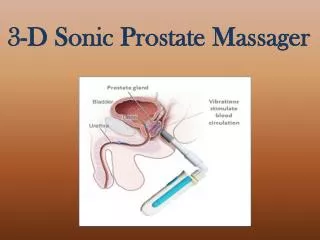
Shear Wave Imaging • Measurement of elasticity is expressed as Young’s Modulus (kPa) and displayed on a color scale • In the areas without color, the shear wave is not generated and therefore the elasticity is not measured
Advantages of Shear Wave • Patient presented with a hypoechoic nodule on US (black lesion with arrow) • SWE was performed and the nodule was found to be of low stiffness (<20kPa). Note the blue area with the yellow arrow. • However, another lesion was found with a high stiffness (75kPa) and was found upon biopsy to be a Gleason grade 7 prostate cancer. Note the red lesion with the red arrow.
Results • A total of 32 patients participated in the study, providing 192 sextants • Average patient age was 64.2 years (range 53-79) • Average PSA was 5.05 (range 0.21-18.6). A total of 14 foci of cancer were detected in 7 of the 32 patients (22%)
Results • Based on the SWE findings, 173/178 sextants were true negatives • 14/14 sextants were true positives • There were 5 false positives and 0 false negatives. This produced a sensitivity of 100%, specificity of 97%, positive predictive value of 74%, and negative predictive value of 100%
Benign Prostate • Recent elevation of PSA from 8 to 12.3 • Homogeneous blue reading indicates uniform low stiffness (< 20kPa), likely ruling out carcinoma
True Positive (Gleason 6 CA) • Low PSA but firm left side on DRE • B-mode imaging and Color Doppler unremarkable • Shear wave elastography demonstrates a posterior high stiffness lesion on the left side, measuring 119kPa • Confirmed on biopsy to be a Gleason 6 carcinoma
False Positive • Stiff (73kPa) and echogenic lesion that suggested malignancy. Note the posterior red lesion circled. • Upon biopsy it was found to be benign prostatic tissue that had been calcified • Current limitation of SWE
False Positive • High (67kPa) stiffness nodule noted on SWE. Note the posterior yellow-orange area circled. • Found on biopsy to be inflamed prostatic tissue with focal calcifications
Conclusion • Transrectal shear wave elastography of the prostate has the potential to become the primary imaging modality for detection of prostate cancer • With a high negative predictive value, this technique could eliminate a significant number of biopsies
References • [1] Bercoff J. “ShearWave Elastography.” Supersonic Imagine White Paper. October 2008. • [2] Correas JM, Khairoune A, Tissier AM, et al. “Trans-rectal quantitative shear wave elastography: application to prostate cancer – a feasibility study.” Proceedings of the European Congress of Radiology, 2011. • Kundra V, et al. Am. J. Roentgenol. 2007;189:830-844 • Kuligowska E, et al. Radiology, September 2001;220, 757-764 • http://www.prostate-cancer.org/pcricms/node/165 • http://gleasonscore.net/