Download
1 / 22
950 likes | 5.01k Views
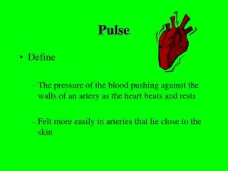
The Arterial Pulse. With each contraction ,the left ventricle ejects a volume of blood into the aorta and on into the arterial tree A pressure wave moves rapidly through the arterial system where it can be felt as the arterial pulse.

E N D
The Arterial Pulse • With each contraction ,the left ventricle ejects a volume of blood into the aorta and on into the arterial tree • A pressure wave moves rapidly through the arterial system where it can be felt as the arterial pulse
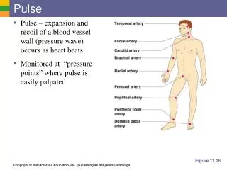
The arterial pulse should be examined in all 4 limbs and both sides of the neck • Radials • Brachials • Carotids • Femorals • Popliteals • Peripheral arteries of the legs :Dorsalis pedis Posterior tibial
How to feel the Pulse • The Radial pulse: • The 3 middle fingers are used • The palmar surface of the fingers overlies the radial A. and encircles the wrist • At first the artery is completely occluded, then gradually release the pressure until maximum feeling of the pulse wave is perceived.
The Carotids • The patient lies down with the head of the bed elevated 30 degrees • Carotid pulsations may be visible just medial to sternomastoid • Place the left thumb on the right carotid A. in the lower third of the neck at the level of the cricoid cartilage, just inside the medial border of the sternomastoid and press posteriorly • Never press both carotids at same time
Brachial Artery • Rest the patient arm with elbow extended palm up • Use the thumb of the opposite hand • Cup your hand under the patient elbow • Feel the pulse just medial to biceps tendon
Femoral Pulse • Press deeply below the inguinal ligament and about mid way between ASIS and SP
Popliteal Pulse • Patient knee should be flexed –leg relaxed • Place the finger tips of both hands so that they meet in the middle line behind the knee and press them deeply in the popliteal fossa
Dorsalis Pedis • Feel the dorsum of the foot just lateral to the extensor tendon of the big toe • If you cannot feel the pulse, explore the dorsum of the foot more laterally
Posterior Tibial Curve your fingers behind and slightly below the medial malleolus of the ankle
Comment on the Pulse • Rate • Rhythm • Volume (amplitude) • Comparison of the two sides • Special character • Condition of the arterial wall
Rate Rate of the pulse at radial artery Normal at rest :60-90 beat / min * if regular: count in 15 sec x 4 * if fast (tachycardia ) or slow (bradycardia) count in 1 min *if irregular count at apex weak beats may not be felt (pulsus deficit)
Rhythm Is the rhythm regular or irregular? If irregular: - Totally irregular (atrial fibrillation) - Irregular beats in a basically regular rhythm (premature beats)
Volume (Amplitude) A--Large amplitude (Bounding pulse) big difference between systolic and diastolic BP * High systolic: increased stroke volume rigidity of aorta * low diastolic :aortic regurge
B--Small amplitude (weak pulse) 1- low stroke volume shock (thready) severe mitral stenosis 2- Aortic stenosis
C--Variation in amplitude 1-pulsus alternans 2-pulsus paradoxus
Comparison of both sides Causes of unequal pulse • Genetic absence or change in the course of the radial artery • Compression of the vessel • Atheromatous plaque • Embolus
Special character • Factors affecting the form • Upstroke (rise) • Duration • Downstroke (fall)
Collapsing and Water hammer pulse • Rapid upstroke • Rapid down stroke • High amplitude • Short duration Found in :Aortic incompetence Hyperdynamic states: Fever Anaemia Thyrotoxicosis
Anacrotic Pulse (Plateau pulse) • Upstroke is slow with a notch on it • Duration of pulse is prolonged • Amplitude is small • In aortic stenosis
Pulsus Bisferiens In combined aortic stenosis and regurge Pulse has 2 peaks: • Upstroke is sharp and rises high to the first peak • Falls and rises again to a second peak A double pulse is felt and seen in the carotid