Download

1 / 68
730 likes | 1.2k Views
VIRAL HEPATITIS. Hepatitis viruses. Dr M Aamir Mirza MBBS,MCPS,M.Phil,Ph.D MABCP(USA),CS Path(UAE). VIRAL HEPATITIS. Viral hepatitis is an acute inflammation of the liver resulting in a clinical illness ,characterised by: A prodromal phase. Fever. Jaundice.

E N D
VIRAL HEPATITIS. Hepatitis viruses. Dr M Aamir Mirza MBBS,MCPS,M.Phil,Ph.D MABCP(USA),CS Path(UAE)
VIRAL HEPATITIS. • Viral hepatitis is an acute inflammation of the liver resulting in a clinical illness ,characterised by: • A prodromal phase. • Fever. • Jaundice. • Symptoms related to GIT e.g nausea and vomiting.
CLINICO PATHOLOGICAL EVENTS. • Regardless of the type of virus as a causative agent, histopathological symptoms are observed in the liver. • After entering the blood the virus infects hepatocytes. • A diffuse inflammatory reaction occurs in the lobules resulting in disruption of liver cell cords.
Spotty parenchymal degeneration of hepatocytes occurs followed by necrosis. • Reticuloendothelial (kupffer cell) hyperplasia occurs with periportal infiltration by mononuclear cells. • Localised areas of necrosis with ballooning and acidophillic bodies are observed frequently.
Macrophages containing lipofuscin accumulate near degenerating hepatocytes. • Bile canaliculi become disrupted resulting in blockage of biliary excretion following liver enlargement or necrosis. • The damaged liver tissue is generally restored in 8-12 weeks.
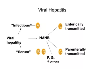
HEPATITIS VIRUSES. • Viral hepatitis is a systemic disease that primarily involves the liver. • The major viral pathogens causing hepatitis are: • Hepatitis A virus (infectious hepatitis). • Hepatitis B virus (serum hepatitis) • Hepatitis C virus (post transfusion hepatitis) • Hepatitis D virus (delta agent) • Hepatitis E virus (enteric hepatitis) • Hepatitis G virus (post transfusion).
HEPATITIS A VIRUS. • It is a member of Picorna viridiae family. • Contains a linear single stranded RNA. • Route of infection: fecal –oral. • Common in children and young adults. • Starts with a prodromal phase followed by jaundice.
Viraemia occurs 2 weeks before and remains for more than 1 week after jaundice. • Virus appears in the stool 2 weeks before and remains for 2 weeks more after jaundice. • The virus is rarely isolated from urine.
Relapses may occur 1-4 months after resolution of initial symptoms . • Hepatitis A is widespread worldwide. • Outbreaks are common in families,institutions,summer camps,day care centers,neonatal ICUs and military troops. • Recurrent epidemics are a permanent feature.
Large outbreaks have been attributed to consumption of infected oysters and clams. A large outbreak had erupted d/t consumption of frozen strawberries in the USA during 1997.
HEPATITIS B VIRUS. • It is worldwide in distribution. • There is no seasonal trend. • There are certain well defined risk groups such as: • Parenteral drug abusers. • Health care personnel. • Recipients of multiple transfusions.
High risk groups. • Cases of haemodialysis. • Cases of organ transplants. • Newborns of mothers with hepatitis B. • Screening of blood donors has been made mandatory for hepatitis B, due to which the number of new cases has declined considerably.
COMMON SOURCES OF INFECTION. • Infected/improperly sterilized syringes and needles, scalpels,blades,tattooing and piercing instruments. • The virus surface antigen can be detected in • Saliva, nasopharyngeal washings, semen, menstrual fluid, vaginal secretions and blood.
TRANSMISSION. • The infection is transmitted through close contact by oral, sexual or other intimate exposure. • Asymptomatic carriers are important sources of transmission. Estimated ratio of anicteric to icteric cases is 4:1.
SOURCES OF HEPATITIS B INFECTION HBV SOURCES OF HEPATITIS B INFECTION HBV HBV
STRUCTURE OF HBV. • Electron microscopy of HBsAg positive serum reveals 3 morphological forms. • 1-Spherical particles 22 nm in diameter. This is the most numerous form which is made up exclusively of HBsAg.
2-Tubularfilamentous form: Has same diameter but may be 200nm long. These forms are produced from over production of HBsAg. • 3-Larger 42 nm spherical virions originally called Dane particles. These are observed less frequently.
The outer surface envelope contains HBsAg. It surrounds the inner nucleocapsid core that contains HBcAg. • The particles containing HbsAg are complex. Each contains: • Group specific antigen a • Mutually exclusive sub determinants(2 pairs)d/y and w/r.
There are 4 phenotypes • Adw,ayw,adr and ayr. • These phenotypes are used as virus specific markers that have epidemiological significance.
VIRAL GENOME. • The viral genome is partially double stranded circular DNA . • Member ofHepadnaviridae family • The virus produces genomic DNA by reverse transcription with mRNA as a template.
PATHOGENESIS. • The virus resists heat at 37degree C for 60 minutes and remains viable after drying for at least 1week at 25 degree C. • HBV but not HBsAg is sensitive to high temp.(100 degrees C). Sodium hypochlorite destroys antigenecity within 3 minutes.It is not destroyed by UV irradiation.
PATHOGENESIS. • Exposure to HBV leads to infection commonly known as Serum Hepatitis. • During the initial phase high concentration of HBV may be present in the blood and communicability is highest.
PATHOGENESIS. • The virus infects hepatocytes and the surface antigen is represented on the surface of cells. • Cytotoxic T lymphocytes mediate an immune attack against the viral antigen followed by inflammation and necrosis. • Cell mediated immune injury occurs .
The virus does not produce any cytopathic effect. • Ag-Ab complexes result in : • Arthralgias. • Arthritis. • Urticaria, • Glomerulonephritis. • Vasculitis. • Cryoglobulinaemia.
HBV genome has no oncogen but the persistent cell regeneration in attempt to replace the dead hepatocytes is the cause of hepatocellular carcinoma. • Malignant transformation may also arise d/t insertional mutagenesis as a result of integration of HB genome into the hepatocyte DNA. This integration may lead to uncontrolled growth d/t activation of a cellular oncogene.
In cases of HBV a cobblestone appearance characterised by swollen and pale hepatocytes is seen which is more common in asymptomatic carriers. • It has a favorable prognosis as it usually does not progress towards cirrhosis.
OUTCOME OF HBV INFECTION. HBsAg persists throughout the clinical course of the disease and typically disappears by the 6th month after exposure. 1-The disease may resolve. 2-Carrier stage may develop. 3- Fulminant hepatocellular necrosis.
CHRONIC CARRIER STAGE: • When HBsAg persists for more than 6 months in the presence of HBeAg or HBe the patient goes into a chronic carrier stage in which HBsAg persists for many years.
PATHOGENESIS OF CARRIER STAGE. • During viral replication some of the progeny DNA integrates into the host cell genome and maintains the carrier stage. • Small amounts of viral DNA are also detectable.
FULMINANT HEPATOCELLULAR NECROSIS. • This condition is seen in 1-2% of cases withHBV infection. • The histological changes range from inflammation and necrosis to collapseof the normal reticulum framework with bridging b/wportal triads and terminal hepatic veins.
HEPATITIS C VIRUS. • It is a member of flavi virus family. • Infections are prevalent throughout the world. • It has at least 6 genotypes. The genetic variation results in a hypervariable regionin the envelope glycoprotein.
The mutation rate is high in the envelope gene coupled with the absence of a proofreading function in the virion encoded RNA polymerase. As a result multiple subspecies occur in the blood of an infected person.
TRANSMISSION AND PATHOGENESIS. • HCV is one of the most prevalent blood borne pathogens and is transmitted by direct percutaneous exposure to infected blood/blood products. • It resembles HBV in its pathogenesis. • Infection results in death of hepatocytes d/t cytotoxicity of T cells andstrongly predisposes to hepatocellular carcinoma.
The rate of chronic carriage is higher than that of HBV and alcoholism is a strong predisposing factor to hepatocellular carcinoma.
HEPATITIS D VIRUS. • Also called Delta virus. • It is a defective virus which can not replicate by itself as it lacks the genes for it’s envelope protein. • It uses the surface Ag of HBV as it’s envelope protein and HBV acts as a helper virus for HDV.
STRUCTRE. • HDV is an enveloped virus with a single stranded RNA genome with –ve polarity & a covalently closed circle. • The RNA genome is very small & codes only one protein, which is the internal core protein called delta Ag. • HDV has no virion polymerase.
The host cell RNA polymerase transcribes the genome RNA & helps in its replication. • HDV genome is a ribozyme (has the ability to self cleave and self ligate during replication. • HDV replicates in the nucleus of host cell.
TRANSMISSION AND PATHOGENESIS. • HDV has only one serotype. • It is transmitted through the same routes as those for HBV. • Hepatocyte damage occurs dt T cell cytotoxicity. • Patient get co-infected or super-infected by HDV.
Super infection is more severe resulting in a fulminant and life threatening hepatitis,chronic hepatitis or liver failure.
HEPATITIS E VIRUS. • A major cause of enterically transmitted hepatitis. • Occurs in epidemic forms in developing countries d/t faecally contaminated water supplies. • A high mortality rate has been observed in pregnant women. • Animal strains are seen in rodents & cattle.
STRUCTURE AND PATHOGENESIS. • HEV is a non enveloped single stranded RNA virus and a member of calicivirus family (recently placed in it’s own genus; the hepevirus). • The disease resembles HAV. • Does not result in chronic liver disease or carrier stage.
HEPATITIS G VIRUS. • Isolated in 1996,from a patient o post transfusion hepatitis. • It is a member of flavi virus family as is HCV. • It causes a chronic infection lasting for decades. • It is found to be transmitted through blood & sexual contact.
LABORATORY DIAGNOSIS OF VIRAL HEPATITIS. • 1-CLINICAL SIGNS & SYMPTOMS. • 2-BIOCHIMICAL TESTS (LFTs). • 3-HISTOPATHOLOGY. • 4-IMMUNOGLOBULIN LEVELS. • ELEVATED • HAV can be detected in blood & stool, by immunoassays.