Download
1 / 24
310 likes | 678 Views
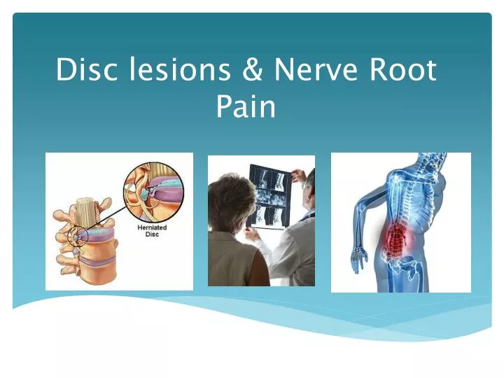
Disc lesions & Nerve Root Pain. Definition of Disc lesion. Lesion is a non-specific term used in medicine to refer to a pathology of tissue. It indicates an area of tissue that has been injured, destroyed, altered (for the worse) or has a problem. Disc lesion refers to disc degeneration

E N D
Definition of Disc lesion Lesion is a non-specific term used in medicine to refer to a pathology of tissue. It indicates an area of tissue that has been injured, destroyed, altered (for the worse) or has a problem. Disc lesion refers to disc degeneration Leads to reduced water content and therefore reduced shock absorption Leads to nucleus pulposes collapsing through annulus fibrosis to adjacent vertebra (known as Schmorl’s nodules)
Causes • Trauma • Flexion rotation injuries- heavy object is lifted (gives rise to tear of the posterior longitudinal ligament and results in bulging of the disc) • Degeneration • Disc loses elasticity due to collagen changes and decrease in water content results in inability to handle compression forces • Increased pressure • Nucleus absorbs moisture during physical illness or emotional stress, swells and presses against annulus fibrosis
Risk factors Age: Most common in middle age, especially between 35 and 45, due to aging-related degeneration of the discs. Weight: excess body weight causes extra stress on the disks in your lower back. Occupation: People with physically demanding jobs have a greater risk of back problems. Repetitive lifting, pulling, pushing, bending sideways and twisting also may increase your risk of a herniated disk. Working night shift has also been found to increase your risk. Pregnancy
Pathology Changes start asymptomatically but causes weak link Most injuries occur at L4,5 and L5, S1 Prolapse to anterior or lateral causes osteophytes which later attach to each other, resulting in loss of movement Posterior prolapse causes more problems Disc itself or osteophytes exert pressure on spinal cord or nerve roots
Symptoms Sudden pain when picking up heavy object- initially slight but worsens and can impair movement. Repeated attacks occur suddenly e.g. sneezing or coughing Pain after a prolonged sustained position Central or referred symptoms- not always clearly defined Proximal worse than distal Pain diminishes when lying down with knees supported or hanging in a specific position
Signs Young, healthy patient Lateral tilt of the pelvis Increased lumbar lordosis Gluteal area sensitive to palpation Protective muscle spasm Sitting, coughing and sneezing painful Decreased intervertebral movements
X-Rays AP- Sometimes shows tilt of vertebra Lateral- Narrowed disc space
Treatment • OMT • Rotation (grade 4-) • Longitudinal • Static traction • Palpation techniques (except when ext. comparable sign) • Electrotherapy • Exercises • Abdominal stabilisation • Strengthening quadriceps and gluteal mm. • Neural mobilisation • Posture correction and kinetic handling • Surgery • http://www.youtube.com/watch?feature=player_embedded&v=i6r5ivym8Ug
Advice Avoid sitting positions (driving or bathing) Sit with knees lower than hips and use lumbar cushion Avoid sustained positions Avoid rotation movements when picking up objects Avoid sudden, jerky movements (sporting activities) Do not pick up heavy objects Swimming is good exercise- strengthens erector spinae Wear a brace during activities which aggravate backache
Nerve Root Pain Pressure on the nerve root causing pain Pain not from nerve itself but as result from venous congestion First sign is pins and needles in distal region of affected dermatome, pain intensifies and arterial blood circulation restricted Nerve conduction suspended and nerve fall-out develops
Degree of impairment • Depends on: • Strength of initial impulse • Duration of abnormal pressure (longer, the worse it becomes)
Causes Disc prolapse Disc protrusion Osteophytes Traction injuries Swelling in intervertebral canal Stenosis Deep-seated muscle spasms Hypertrophic capsule
Physical manifestations • Nerve root irritation: • Increased reflexes • Abnormal sensation or paraesthesia • Nerve root pressure: • Decreased reflexes • Loss/ no sensation • Muscle weakness- long term atrophy
Manifestations cont. • Pressure on spinal cord: • Gait disturbances • Bilateral pins and needles • Bladder dysfunction • Increased reflexes below level of lesion • Clonus and Babinski (+) • Pressure on caudaequina • Saddle anaesthesia • Urine retention
Characteristics of nerve root pain • Area: • Well defined throughout dermatome or dominates distal part of dermatome • Nature: • Severe pain, sometimes total loss of function, may be latent, often undulent and builds up. • Sharp, shooting pain which may paralyse patient • Root pain of C7 refers to medial border of scapula with cervical movements (Cloward areas) • S1 refers to medial buttock • Acute phase, pain severe, sub-acute and chronic, pain is intermittent
Movement: • Either distal or latent pain in distal segment • More distal pain is caused, more careful the management • Deformities: • Protective deformities occur • Patient stands on one leg with other leg bent and toes resting on the floor
Treatment • Severe nerve root pain: • Hospital traction or traction as out patient • (Neurological examination is an ABSOLUTE prerequisite, palpation techniques are CONTA-indicated) • Steroid injections • As soon as symptoms improve, mobilisation techniques (Gr. 4) may be added to traction • Treatment suspended after 85% improvement • If symptoms extremely severe, surgery is indicated
Specific treatment • Cervical root pain: • Constant traction • Collar for support to restrict movement • Advice regarding sleeping positions • Lumbar root pain: • Complete bed-rest • Constant traction • Takes longer to react to treatment
Chronic and intermittent nerve root pain • Chronic: • History of prolonged pain in back and leg • Strong treatment techniques indicated (e.g traction and SLR) • Intermittent: • Pain only occurring in one dermatome e.g the knee • Local hypertrophy and palpation tenderness of interspinal ligament • Quick active tests and palpation techniques do not reproduce symptoms • Treatment: Trigger points Neural mobilisation Ultrasound
Effectiveness of therapeutic lumbar transforaminal epidural steroid injections in managing lumbar spinal pain. OBJECTIVE: To evaluate the effect of therapeutic transforaminal lumbar epidural steroid injections in managing low back and lower extremity pain. OUTCOME MEASURES: The primary outcome measure was pain relief (short-term relief = up to 6 months and long-term > 6 months). Secondary outcome measures were improvement in functional status, psychological status, return to work, and reduction in opioid intake.
RESULTS: 27 studies met inclusion criteria, 15 randomized trials and 10 non-randomized studies. For lumbar disc herniation, the evidence is good for transforaminal epidural with local anesthetic and steroids, whereas it was fair for local anesthetics alone and the ability of transforaminal epidural injections to prevent surgery. For spinal stenosis, the available evidence is fair for local anesthetic and steroids. The evidence for axial low back pain and post lumbar surgery syndrome is poor, inadequate, limited, or unavailable. CONCLUSION: In summary, the evidence is good for radiculitis secondary to disc herniation with local anesthetics and steroids and fair with local anesthetic only. Evidence is limited for axial pain and post surgery syndrome using local anesthetic with or without steroids.
References Barnes,R.2011.NEUROMUSCULAR-SKELETAL REHABILITATION DICTATE.(Unpublished dictate.) University of the Free State , Free State. Manchikanti L, Buenaventura RM, Manchikanti KN, Ruan X, Gupta S, Smith HS, Christo PJ. 2012. Effectiveness of therapeutic lumbar transforaminal epidural steroid injections in managing lumbar spinal pain.(http://www.ncbi.nlm.nih.gov/pubmed/22622912) Retrieved 22 August 2012 South Wales Osteopathic Society. Disc lesions. (http://osteopathywales.com/index.php?option=com_content&view=article&id=204:disc-lesions&catid=22:medical-conditions) Retrieved 19 August 2012.
References cont. Asher, A. 2006. Lesion (http://backandneck.about.com/od/l/g/lesion.htm). Retrieved 19 August 2012. Health on Care. 2012. Disc Herniation, Prolapse Disc : Definition, Causes, Symptoms, Diagnosis, Prevent and Treatment (http://www.healthoncare.com/disc-herniation-prolapse-disc.html). Retrieved 19 August 2012.