Download
1 / 16
160 likes | 329 Views
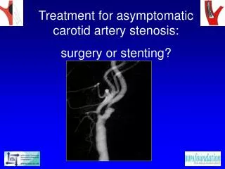
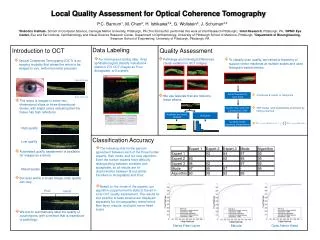
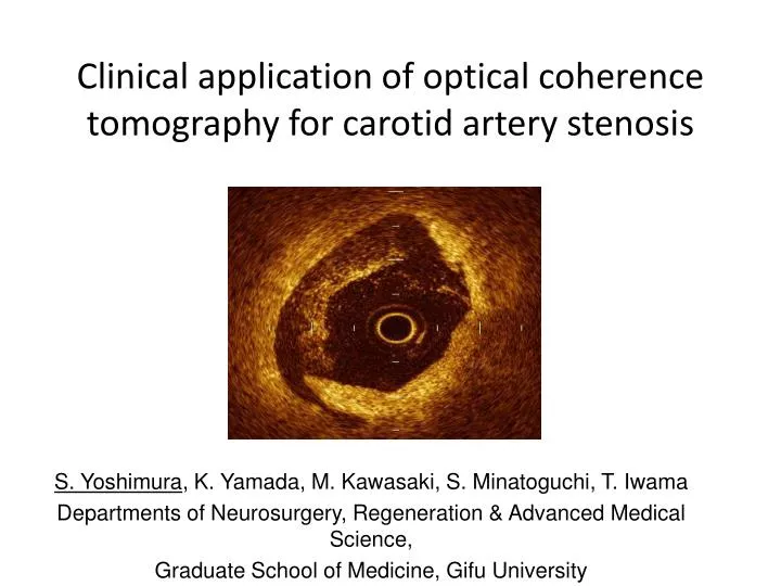
Clinical application of optical coherence tomography for carotid artery stenosis. S . Yoshimura , K . Yamada, M . Kawasaki, S . Minatoguchi , T . Iwama Departments of Neurosurgery , Regeneration & Advanced Medical Science, Graduate School of Medicine, Gifu University. Introduction.

E N D
Clinical application of optical coherence tomography for carotid artery stenosis S. Yoshimura,K. Yamada,M. Kawasaki,S. Minatoguchi,T. Iwama Departments of Neurosurgery,Regeneration & Advanced Medical Science, Graduate School of Medicine, Gifu University
Introduction Intravascular optical coherence tomography (OCT) is a non-contact, light-based high-resolution imaging device for plaque characterization, which provides additional morphological information beyond intravascular ultrasound (IVUS) images. OCT Imaging System OCT (Image Wire, Light-lab Imaging, Goodman, Co, Ltd, Nagoya)
Methods probe interface unit with auto pull-back system Guardwire for ECA occlusion Since OCT is approved only for coronary arteries in Japan, its use for human carotid arteries was approved by our institutional ethics committee (No. 21-108), and the study was registered on the internet (UMIN 000002808). 9 Fr guiding catheter with a balloon for CCA occlusion
Case 1 83 M The patient presented with temporary lt. hemiparesis. rt. CAG Just before stenting, plaque shape was changed. So, we performed OCT and IVUS first.
Case 1 OCT IVUS black blood MRI HE staining
Intraluminal findings Thrombus VH-IVUS OCT IVUS Flap formation Ulceration
Intraplaque findings Fibrous VH-IVUS OCT IVUS Calcification Lipid
Postprocedural findings Plaque protrusion Endotheliali- zation
Discussion • I this study, OCT examination was performed safely without any complication. • The typical OCT image has an axial resolution of 10 µm, which is 10 times higher than those of other clinical imaging. • Thrombus, fibrous cap rupture and postoperative plaque protrusion could be detected by OCT examination, which were hard to be obtained by other methods.
Conclusions: • The present study suggested that OCT might be useful to investigate the carotid artery stenosis. • One of the important limitations of OCT is degree of tissue penetration. • Then, combination of OCT and VH-IVUS may be a good way to evaluate total plaque morphology.