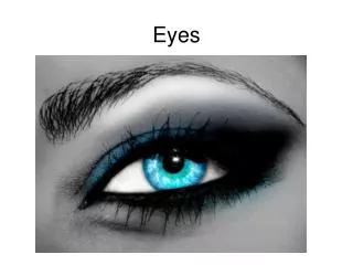
Eyes
Eyes. Dr Bruce Davies www.bradfordvts.co.uk. You are not alone!. A very popular topic How much time at medical school? What do the acuity numbers mean!. Special history. One or both? What disturbance of vision? Rate of onset? Any blind spots?


Eyes
E N D
Presentation Transcript
Eyes Dr Bruce Davies www.bradfordvts.co.uk
You are not alone! • A very popular topic • How much time at medical school? • What do the acuity numbers mean!
Special history • One or both? • What disturbance of vision? • Rate of onset? • Any blind spots? • Any associated symptoms e.g. floaters? flashing lights? • Exactly what is worrying the patient.
Contact lens use? • Myopia? (increases risk of retinal detachment 10 fold) • Any family history? (FH of glaucoma in a 1st degree relative gives you a 1/10 lifetime risk, or squint) • Any history of diabetes, hypertension or connective tissue disease?
Examination • Snellan chart, 3m or 6m, simple text for near vision, • Pinholes • Fields, remember red and the quality of the red, simple 4 quadrant testing. • Pupils: a bright torch and magnifying glass • Squint • Movements • Opthalmoscopy: Start at 10, red reflex?, green filter enhances blood vessels, dilate prn, risk of acute closed angle glaucoma remote.
Clinical classification • Red eye • Lids and tears • Slow visual loss in the quiet eye • Trauma • Squints, new and congenital, rare movement disorders • …..(then a rare specialist rag bag)
Red eye Conjunctivitis Commonest, an uncomfortable red eye. Bacterial • Discomfort. Purulent discharge. Spreads from one eye to the other. Vision normal. Uniform engorgement Chloramphenicol first choice (?)
Conjunctivitis Viral • Often with an URTI. Gritty. Discomfort. Watery discharge. May last many weeks. • Photophobia. Small corneal opacities may develop. Prolonged (often adenoviral) may need specialist therapy with steroids. Chloramphenicol to prevent 2nd infection.
Conjunctivitis Chlamydia • Mucopurulent, cornea inflamed, visual loss. Often with STD. Permanent damage possible, topical and? systemic tetracyclines. Refer. Infants • Less than one month is notifiable disease - any cause. May lead to scarring and permanent damage. Refer most. Allergic • Itching and discomfort. Chemosis and visual acuity loss possible. Papillae and if big cobblestones. Cromoglycate may take days to start to work if bad.
Episcleritis / scleritis Red sore eye. No discharge. Localised (viz. conjunctivitis=generalised) inflammation. • Episcleritis usually self limiting and idiopathic, no treatment needed. • Scleritis often with CT diseases, dangerous (perforation possible) Refer.
Corneal ulcers • Any infection, Abrasion, topical steroids, contact lens use. • PAIN. - Except zoster • May be general or localised inflammation. • Must stain. Should evert upper lid to exclude a sub tarsal FB • ?Hypopyon - pus in anterior chamber. • Refer most (except small abrasions - but refer if big or longer than 36 hours) • Remember recurrent abrasion syndrome.
Anterior uveitis • The uveal tract. So iritis, iridocyclitis and anterior uveitis are synonyms. • At risk: HLA-B27, CT diseases, past attacks, juvenile arthritis, sarcoid. • PAIN, then photophobia then visual loss. • Ciliary flush. As it gets worse the pupil gets small and reactions get sluggish, hypopyon, keratitis (back of cornea). These markers of it getting worse are bad news. • Refer all.
Acute closed angle glaucoma • Often starts in the evening. Especially in those over 50 years. • Severe pain first. Impaired vision and haloes around lights. May have history of past episodes relieved by going to sleep (the pupil constricts during sleep). • Refer even if attack spontaneously resolves.
Lids and tears Chalazion • = meibomnian cyst. In the lid. Warm compresses and chloramphenicol. Persistent - incise. • Recurrent: ? DM, ? blepharitis, ? roseacea. • Can cause astigmatism from pressure.
Stye • An infection of lash follicle. May be head of pus - nick with needle. Or warm compresses and chloramphenicol.
Marginal cysts • Non infected cysts from sweat or sebaceous lid glands, if a problem can often be simply treated with a nick with a needle - small.
Blepharitis • Common, underdiagnosed. Persistently sore eyes. Gritty. Often with chalazions or styes. Inflamed lid margins, crusts, may have inflamed lids. • Associated with psoriasis, eczema and roseacea. • Keep clean, antibiotic ointment[tetracycline], artificial tears ? oral tetracyclines
Acute dacrocystitis • Medial inflammation over lacrimal sac. Refer, systemic therapy and topical urgently.
Orbital cellulitis • Life threatening and blinding. Usually from sinuses. Especially important in children who may become blind in hours. • Unilateral swollen lids which may not be red. • The patient is ill, there is tenderness over the sinuses, restricted eye movements. ADMIT
Ectropion • Watery eye.. Laxity from age or nerve palsy. Ointment and refer for LA operation to correct. Entropion • Common especially in the elderly. Scarring from the lashes. • Often results from blepharitis or chronic conjunctivitis • Refer
Ingrowing lashes • Damage to lids. May be removed but will often need electrolysis or cryocautery to prevent recurrence.
Watering eyes • Differential diagnosis.- your homework! Dry eyes • Common, • Remember to treat associated blepharitis
Sudden visual loss An easy list really as they all need specialist assessment!
Retinal detachment • Floaters, photopsias, the shadow or curtain across the sight. Optic neuritis • More women, pain on moving the eye, central scotoma Posterior vitreous detachment • Aged 50+, flashing lights, floaters Vitreous haemorrhage • Floaters, red haze may be present. Red reflex absent.0
Disciform macular degeneration • Sudden disturbance of central vision. • Vascular occlusions • Field loss. Diabetes, hypertension • Migraine • Youth, headache, zigzag lines, multicoloured lights. • Cerebrovascular disease • Elderly, bilateral loss.
Slow visual loss Refer to optician then ? refer. • Cataracts • Corneal opacities • Macular problems • Retinal problems
Trauma • Refer ! • Unless really trivial
Squints • Refer • Remember the orthoptist • Can you do a cover test?