Download
1 / 50
500 likes | 686 Views
The electrolytes cassette. The electrolytes cassette. An electrolyte is the ionized (or ionizable) constituents of a living cell, blood or other organic matter when ionized it carries a net electrical charge

E N D
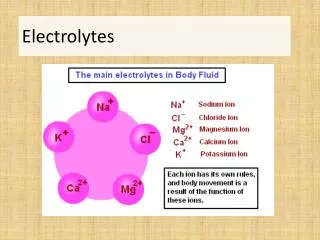
The electrolytes cassette • An electrolyte • is the ionized (or ionizable) constituents of a living cell, blood or other organic matter • when ionized it carries a net electrical charge • sodium(Na+), potassium(K+), calcium(Ca++), magnesium, chloride(Cl-), phosphate, and bicarbonate (HCO3-)….
The electrolytes cassette • An electrolyte • is the ionized (or ionizable) constituents of a living cell, blood or other organic matter • when ionized it carries a net electrical charge • sodium(Na+), potassium(K+), calcium(Ca++), magnesium, chloride(Cl-), phosphate, and bicarbonate (HCO3-)….
Intracellular fluid Extracellular fluid Why do vets care? • All higher life forms require a subtle and complex electrolyte balance between the intracellular and extracellular environments Na+ 10 mmol/L K+ 145 mmol/L Na+ 140 mmol/L K+ 4 mmol/L
Why do vets care? Na+÷ K+ 142 ÷ 6.5 = 22 !!! • Serious electrolyte disturbances may lead to cardiac and neurological complications, and most are medical emergencies • Knowledge of blood electrolyte concentrations may help to make a diagnosis or dramatically influence treatment
Why do vets care? • Fluids lost from the animal have an unknown concentration of electrolytes • Fluids added to the patient have a known concentration but an unknown effect • Drugs given to the patient can dramatically alter the concentration of electrolytes
K+ (+) (-) K+ K+ Potassium • Just like a ‘battery’ some cells within the body have a voltage • -ve inside the cell membrane • +ve outside the cell membrane • The concentration of potassium in the extracellular fluid can dramatically alter this voltage • Important role in the automaticity of the heart and transmission of signals in the nerves
How is potassium balance maintained? FOOD PLASMA K+ ↔ CELLS 90% URINE FAECES SWEAT
INSULIN INSULIN 3Na+ 3Na+ + + 2K+ 2K+ 2K+ + 3Na+ INSULIN Intracellular fluid Extracellular fluid
HYPERKALAEMIA HYPONATRAEMIA + + ADRENAL GLANDS ALDOSTERONE + Na+ KIDNEY K+ THIS FLUID BECOMES URINE
+ ADRENAL GLANDS Na+÷ K+ 142 ÷ 6.5 = 22 !!! ALDOSTERONE + Na+ KIDNEY K+ THIS FLUID BECOMES URINE
Elevated blood potassium Hyperkalaemia • Long term hyperkalaemia uncommon if renal function normal • Increased oral intake is an unlikely cause • Clinical manifestations reflect changes in cell membrane voltage • muscle weakness • changed electrocardiogram (ECG)
Hyperkalaemia • ECG changes • Some vets consider they do not need to measure potassium if they have an ECG machine • The waveform cannot be used to predict a plasma potassium concentration 1 2 3 4
Causes of hyperkalaemia • Urinary bladder rupture • Urinary tract obstruction • treatment must focus on removing obstruction and restoring urine flow • Kidney failure
Causes of hyperkalaemia • Drugs • Some diuretics • ACE inhibitors (commonly used during heart failure) • Iatrogenic (“vet induced”) • potassium-rich fluid therapy (‘drips’) • excessive oral potassium supplementation
Causes of hyperkalaemia • Significant tissue destruction • Massive amounts of intracellular potassium released into the circulation • Chemotherapy • Severe trauma
K+ H+ Causes of hyperkalaemia • Metabolic acidosis • Intracellular translocation of hydrogen ions
Treatment of hyperkalaemia • Mild (5.9 to 6.4 in dogs, 4.6 to 6.4mmol/L in cats) • intravenous fluids with low potassium • Moderate (6.5 to 7.5 mmol/L) • as above with insulin • Severe (>7.6 mmol/L) • calcium gluconate or • sodium bicarbonate
Clinical features of hypokalaemia • May have no clinical signs • not usually apparent until serum K+ < 3mmol/L • Muscle weakness • Cardiac muscle dysfunction (arrhythmias)
Causes of hypokalaemia • Iatrogenic causes • some diuretics • aggressive IVFT • excessive insulin • Chronic vomiting • Chronic kidney failure • Monitor in any patient that is: • NOT eating • receiving intravenous fluid therapy
Cats and hypokalaemia • Cats seem particularly prone to developing hypokalaemia • Many cats receiving standard intravenous fluids benefit from K+ supplementation • Cats with chronic renal failure may benefit from oral supplementation
Useful tips when measuring [K+] • Tourniquet released after maximum of one minute to avoid venous stasis • haemoconcentration and drives potassium out of cells • Avoid ‘fighting’ with patient • Analysis without delay
Causes of hyperkalaemia • Laboratory artefact: occurs either during or after sampling • NOT a problem for the animal • haemolysed blood sample (especially puppies and Akita dogs) • leucocytosis • thrombocytosis • Tri-K EDTA
Case example: ketoacidotic cats • A cause of hyperkalaemia AND hypokalaemia • Body K depleted • polyuria and insulin deficiency, vomiting • Ketoacidotic patients • in general acidosis is associated with movement of K+ ions from ICF to ECF • rapidly reversed when insulin therapy and IVFT commenced • Potassium monitoring is vital
Disorders of sodium and water • Volume and concentration of body fluids are maintained within a narrow range by regulation of sodium and water loss • The kidney plays a crucial role: balancing the excretion of salt and water with their intake Extracellular Intracellular
We start with a bucket of water… This membrane will let water pass but not electrolytes
Intracellular fluid Extracellular fluid
Blood sodium concentrations • Indication of the amount of sodium relative to the amount of water in the ECF • Provides no information about the total body sodium • Not an indicator of dehydration – the vet must use other signs
Dehydration • Loss of body water • Loss will occur from the ECF • Fluid lost from the body will have an unknown concentration of electrolytes ECF OSMOSIS ICF
What is the point? • Serum sodium concentrations do not help the vet decide if the patient is dehydrated • A knowledge the patient’s hydration status and sodium concentration will help the vet to decide what the likely underlying mechanism was and how best to provide intravenous fluid therapy
Normovolaemia Hypervolaemia Hypovolaemia Pure water deficit Hypotonic loss Gain of sodium Diabetes inspidus Fever High environmental temp Inadequate access to water Salt poisoning Hypertonic IVFT Hyperaldosteronism Diuresis e.g. frusemide Chronic renal failure Vomiting Diarrhoea Burns Third space loss Increased serum sodium concentration Evaluate volume status
Do not change the plasma sodium too quickly • A change in total brain water of >10% is incompatible with life • smaller changes associated with neurological symptoms • Organic osmolytes • Severe neurological consequences if serum sodium concentration changed too quickly • Serum sodium concentration should be monitored serially • change of < 0.5 mmol/L/hour ECF OSMOSIS ICF
Plasma Na+ K+ Frusemide Spironolactone ACEi Case example: congestive heart failure • Diagnosed with congestive heart failure three years ago • ‘Accumulated’ drugs over that time: frusemide, enalopril and spironolactone • Presented collapsed with neurological signs
When to test • Any patient that presents in an emergency • Any sick patient • Any patient receiving intravenous fluid therapy (particularly those receiving large volumes quickly) • Any patient that isn’t eating
When to test • Any sick patient prior to anaesthesia • Any patients • receiving multiple medications for heart failure • with chronic renal failure • receiving potassium supplementation • Any patient worth taking blood from?
Question your (reluctant) vet? • How do you know if your diabetic crisis cats are hypokalaemic or hyperkalaemic? • Most cats benefit from having potassium added to their intravenous fluids; how do you know how much to add? • How are you going to deal with your next case that might have Addison’s disease?
Question your (reluctant) vet? • If you don’t know what type of dehydration you have, can you reliably pick the best fluid for your patient? • If your patient has had a chronic hypo- or hypernatraemia, how quickly do you think you can change that without knowing the electrolytes? • Did you know ‘heart meds’ can lead to electrolyte disturbances?
Question your (reluctant) vet? • Electrolytes can change within the hour; what is the benefit of an out-of-house lab report? • Did you know that electrolyte abnormalities were associated with the following anaesthetic problems? • Low arterial blood pressure • Cardiac arrhythmias and arrest • Delayed recovery
RCVS Practice Standards: ESC • 10.6 Laboratory Facilities • Laboratory facilities for routine diagnostic tests must be available at all times. • This must include electrolytes and blood gases, biochemistry and haematology