Download
1 / 29
290 likes | 399 Views
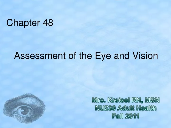
Chapter 48. Assessment of the Eye and Vision. Mrs. Kreisel RN, MSN NU230 Adult Health Fall 2011. Anatomy and Physiology Review. Layers of the eyeball: 3 layers or coats

E N D
Chapter 48 Assessment of the Eye and Vision Mrs. Kreisel RN, MSN NU230 Adult Health Fall 2011
Anatomy and Physiology Review • Layers of the eyeball: 3 layers or coats • Sclera: External Layer: opaque tissue making up the “white” of the eye, and the transparent cornea on the front of the eye. • Uvea: Middle Layer: is heavily pigmented. Consists of the Choroid (Many blood Vessels that supply nutrients to the eye), ciliary body(conets the choroid with the irisand secreates aquous humor), & iris controls amount of light entering the eye (is the colored portion of the external eye in the middle of the pupil). • Retina: innermost layer a thin delicate structure made up of sensory receptors that transmit impluses to the optic nerve.
Anatomy and Physiology Review • Refractive structures and media: Light waves pass through these structors on the way through the eye. Each structorhas different ensity which casues light waves to bend (refract) to some degree to focus on images on the retina. • Cornea: Clear layer that forms external coat on front of the eye • Aqueous Humor: clear watery fluid that fills A&P chambers of eye • Lens: circular convex structure, behind iris, in front to vitreous body, Normal is transparent. Lens bends the rays of light entering pupil so it can be focused on the retina.
Anatomy and Physiology Review • Vitreous Body: clear thick gel that fills the vitreous chamber (space between lens and retina) transmits light and maintains eye shape. • Intraocular Pressure (IOP): Hollow organ, vitreous humor & aqueous humor keep shape and the pressure in the eye. Too low eye collapse, too high called glaucoma
Anatomy and Physiology Review • External structures: • Canthus: place where two eyelids meet in the cornor of the eye. • Conjunctiva: Muscle membranes of the eyes • Lacrimal Gland: upper outer part of each orbit Responsible for tears • Punctum opening at the nasal side of the lid ledges into the lacrimal duct and sac then nose through the nasolacrimal duct. • Muscles, nerves, and blood vessels: • LOOK AT PICTURE 1072
Refraction • Emmetropia—the perfect refraction of the eye • Hyperopia—occurs when the eye does not refract light enough (farsightedness) • Myopia—occurs when the eye overrefracts or overbends the light (nearsightedness) • Astigmatism—a refractive error caused by unevenly curved surfaces on or in the eye, especially of the cornea
Pupillary Constriction • Miosis is pupillary constriction. • Mydriasis is pupillary dilation. • Accommodation is the process of maintaining a clear visual image when the gaze is shifted from a distant to a near object. • Convergence:Turn both eyes inward toward the nose to ensure only a single image of close object is seen
Age-Related Structural Changes • Declining visual acuity: • Decreased eye muscle tone • Ectropion: lower eyelid may relax and fall away fromfrom the eye • Dry eye • Arcus senilis: opque bluish white ring outer edgeof cornea caused by fat deposits • Corneal changes: clarity and shape • Changes in color of sclera • Less ability to dilate pupil • More light needed for reading
Nursing Interventions • Declining visual acuity: • Lie about vision b/c afraid will lose driving privledges • Stigma r/t to blindness • Anxiety • Problems with ADLs and feeling of self worth • Affects job, how support family, life sty;e
Age-Related Functional Changes • Lens yellows. • Accommodation is gradually lost. Lens hardens, shrinks & Loses elasticity and accommodate light decrases. • Near point of vision increases (presbyopia). Hold things farther away to see • Far point decreases. Narrower field of vision • Color perception decreases. • Intraocular pressure increases.
Assessment • Patient history • Nutrition history • Family history and genetic risk • Current health problems
Assessment • Near vision • Visual field • Extraocular muscle function • Color vision
Diagnostic Tests • Imaging assessment • Slit-lamp examination • Corneal staining • Tonometry: measures intraocular pressure Normal Pressure10-21mm hg
Ophthalmoscopy Move toward pts eye from 12-15 inches away and to the side of his/her vision. As you direct the scope at the pupil you should get a red glare (red reflex) means reflection of the light on the retina. If no red glare means lens opacity or cloudy vitreous
Other Diagnostic Tests • Fluorescein angiography: Die in eye, rapid pictures detect retinal circulation deficits • Electroretinography: retina response to light stimulation. Detect blood vessle damage
Chapter 48 NCLEX TIME
Question 1 Which circumstance places the patient at the greatest risk for developing vision disturbances? • History of working with computer • Advanced age • History of diabetes mellitus • Previous employment as a road construction worker
Question 2 What characteristic would the nurse expect to see with age-related changes in an older patient’s eyes? • Yellowing of the sclera • Retinal atrophy • Color blindness • Early-onset glaucoma
Question 3 Which is a priority nursing intervention when providing care to an older patient who has problems with vision? • Review the medication administration record for artificial tears • Review medications before administration • Ensure adequate, nonglare lighting in the patient’s room • Provide written and verbal instruction for nursing education interventions
Question 4 In performing a psychosocial assessment of a patient who has recently experienced vision changes, the nurse should: • Provide the patient with a list of services for the visually impaired. • Meet with family members or significant others to determine if the patient can still perform his ADLs. • Ask the patient how he feels about the changes in his vision and the effectiveness of his coping methods. • Ask the patient if he has made appropriate adjustments in his lifestyle to accommodate his vision changes.
Question 5 What is an appropriate expected outcome for the patient who has undergone an examination of the eye using fluorescein angiography? • Administering mydriatic eye drops for 1 week • Drinking fluids to eliminate the dye • Appearance of bright red–colored urine until the dye is excreted • Staining of the skin for up to 1 week after the test