Chapter 48
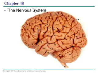
Chapter 48. Nervous Systems. Overview: Command and Control Center. The human brain contains about 100 billion nerve cells, or neurons Each neuron may communicate with thousands of other neurons.


Chapter 48
E N D
Presentation Transcript
Chapter 48 Nervous Systems
Overview: Command and Control Center • The human brain contains about 100 billion nerve cells, or neurons • Each neuron may communicate with thousands of other neurons
Functional magnetic resonance imaging is a technology that can reconstruct a three-dimensional map of brain activity • Brain imaging and other methods reveal that groups of neurons function in specialized circuits dedicated to different tasks
Concept 48.1: Nervous systems consist of circuits of neurons and supporting cells • All animals except sponges have a nervous system • What distinguishes nervous systems of different animal groups is how neurons are organized into circuits
Organization of Nervous Systems • The simplest animals with nervous systems, the cnidarians, have neurons arranged in nerve nets
LE 48-2a Radial nerve Nerve ring Nerve net Hydra (cnidarian) Sea star (echinoderm)
Sea stars have a nerve net in each arm connected by radial nerves to a central nerve ring
LE 48-2b Eyespot Brain Brain Nerve cord Ventral nerve cord Transverse nerve Segmental ganglion Planarian (flatworm) Leech (annelid)
Relatively simple cephalized animals, such as flatworms, have a central nervous system (CNS)
LE 48-2c Ganglia Brain Anterior nerve ring Ventral nerve cord Longitudinal nerve cords Segmental ganglia Insect (arthropod) Chiton (mollusc)
Annelids and arthropods have segmentally arranged clusters of neurons called ganglia • These ganglia connect to the CNS and make up a peripheral nervous system (PNS)
LE 48-2d Brain Spinal cord (dorsal nerve cord) Brain Sensory ganglion Ganglia Squid (mollusc) Salamander (chordate)
Nervous systems in molluscs correlate with lifestyles • Sessile molluscs have simple systems, whereas more complex molluscs have more sophisticated systems
In vertebrates, the central nervous system consists of a brain and dorsal spinal cord • The PNS connects to the CNS
Information Processing • Nervous systems process information in three stages: sensory input, integration, and motor output • These three stages are handled by specialized populations of neurons.
LE 48-3 Sensory input Sensor Integration Motor output Effector Central nervous system (CNS) Peripheral nervous system (PNS)
Sensory neurons transmit information from sensors that detect external stimuli and internal conditions • Sensory information is sent to the CNS, where interneurons integrate the information • Motor output leaves the CNS via motor neurons, which communicate with effector cells • The three stages of information processing are illustrated in the knee-jerk reflex
Sensory neuronstransmit information from sensors that detect external stimuli (light, sound, touch, heat, smell, and taste) and internal conditions (such as blood pressure, blood CO2 level, and muscle tension). • This information is sent to the CNS, where interneurons integrate (analyze and interpret) the sensory input, taking into account the immediate context as well as what has happened in the past. • The greatest complexity in neural circuits exists in the connections between interneurons. • Motor output leaves the CNS via motor neurons, which communicate with effector cells (muscle cells or endocrine cells). The stages of sensory input, integration, and motor output are easiest to study in the relatively simple nerve circuits that produce reflexes, the body′s automatic responses to stimuli. Figure 48.4 diagrams the nerve circuit that underlies the knee–jerk reflex in humans
LE 48-4 Gray matter Cell body of sensory neuron in dorsal root ganglion Quadriceps muscle White matter Hamstring muscle Spinal cord (cross section) Sensory neuron Motor neuron Interneuron
Neuron Structure • Most of a neuron’s organelles are in the cell body • Most neurons have dendrites, highly branched extensions that receive signals from other neurons • The axon is typically a much longer extension that transmits signals to other cells at synapses • Many axons are covered with a myelin sheath
LE 48-5 Dendrites Cell body Nucleus Synapse Signal direction Axon hillock Axon Presynaptic cell Synaptic terminals Myelin sheath Postsynaptic cell
Neurons have a wide variety of shapes that reflect input and output interactions
LE 48-6 Dendrites Axon Cell body Interneurons Sensory neuron Motor neuron
Supporting Cells (Glia) • Glia are essential for structural integrity of the nervous system and for functioning of neurons • Types of glia: astrocytes, radial glia, oligodendrocytes, and Schwann cells
In the CNS, astrocytes provide structural support for neurons and regulate extracellular concentrations of ions and neurotransmitters
Some astrocytes respond to activity in neighboring neurons by facilitating information transfer at those neurons′ synapses. Scientists hypothesize that this facilitation may be part of the cellular mechanism of learning and memory. LE 48-7 50 µm
LE 48-7 Astrocytes adjacent to active neurons also cause nearby blood vessels to dilate, which increases blood flow to the area, enabling the neurons to obtain oxygen and glucose more quickly. 50 µm
. During development, astrocytes induce the formation of tight junctions between cells that line the capillaries in the brain and spinal cord. The result is the blood–brain barrier, which restricts the passage of most substances into the CNS, allowing the extracellular chemical environment of the CNS to be tightly controlled LE 48-7 50 µm
Oligodendrocytes (in the CNS) and Schwann cells (in the PNS) form the myelin sheaths around axons of many vertebrate neurons
Neurons become myelinated during development when Schwann cells or oligodendrocytes growaround axons, wrapping them in many layers of membrane, somewhat like a jelly roll. • These membranes are mostly lipid, which is a poor conductor of electrical currents. • Thus, the myelin sheath provides electrical insulation of the axon. • In the disease multiple sclerosis, myelin sheaths gradually deteriorate, resulting in a progressive loss of body function due to the disruption of nerve signal transmission
In an embryo, radial glia form tracks along which newly formed neurons migrate from the neural tube, the structure that gives rise to the CNS (see Figures 47.14 and 47.15). • Both radial glia and astrocytes can also act as stem cells, generating neurons and other glia. • Researchers view these multipotent precursors as a potential way to replace neurons and glia that are lost to injury or disease, a topic explored in Concept 48.7.
LE 48-8 Nodes of Ranvier Layers of myelin Axon Schwann cell Schwann cell Nucleus of Schwann cell Nodes of Ranvier Axon Myelin sheath 0.1 µm
Concept 48.2: Ion pumps and ion channels maintain the resting potential of a neuron • Across its plasma membrane, every cell has a voltage called a membrane potential • The cell’s inside is negative relative to the outside • Membrane potential of a cell can be measured
LE 48-9 Microelectrode –70 mV Voltage recorder Reference electrode
The Resting Potential • Resting potential is the membrane potential of a neuron that is not transmitting signals • Resting potential depends on ionic gradients across the plasma membrane Animation: Resting Potential
Concentration of Na+ is higher in the extracellular fluid than in the cytosol • The opposite is true for K+
Concentration of Na+ is higher in the extracellular fluid than in the cytosol The opposite is true for K+ LE 48-10 CYTOSOL EXTRACELLULAR FLUID [Na+] 150 mM [Na+] 15 mM [K+] 150 mM [K+] 5 mM [Cl–] 120 mM [Cl–] 10 mM [A–] 100 mM Plasma membrane
The Na+ and K+ gradients are maintained by the sodium–potassium pump (see Figure 7.16). • The fact that the gradients are responsible for the resting potential is shown by a simple experiment: • If the pump is disabled by the addition of a specific poison, the gradients gradually disappear, and so does the resting potential • By modeling a neuron with an artificial membrane, we can better understand resting potential
The sodium–potassium pump: a specific case of active transport. This transport system pumps ions against steep concentration gradients: Sodium ion concentration (represented as [Na+]) is high outside the cell and low inside, while potassium ion concentration ([K+]) is low outside the cell and high inside. The pump oscillates between two conformational states in a pumping cycle that translocates three sodium ions out of the cell for every two potassium ions pumped into the cell. ATP powers the changes in conformation by phosphorylating the transport protein
LE 48-11 –92 mV +62 mV Inner chamber Outer chamber Inner chamber Outer chamber 150 mM NaCl 150 mM KCl 15 mM NaCl 5 mM KCl Cl– K+ Na+ Cl– Sodium channel Potassium channel Artificial membrane Membrane selectively permeable to K+ Membrane selectively permeable to Na+ We start with a model of a mammalian neuron consisting of two chambers separated by an artificial membrane
LE 48-11 –92 mV +62 mV Inner chamber Outer chamber Inner chamber Outer chamber 150 mM NaCl 150 mM KCl 15 mM NaCl 5 mM KCl Cl– K+ Na+ Cl– Sodium channel Potassium channel Artificial membrane Membrane selectively permeable to K+ Membrane selectively permeable to Na+ To produce a concentration gradient for K+ like that of a mammalian neuron, we add 150 mM potassium chloride (KCl) to the inner chamber and 5 mMKCl to the outer chamber.
Like any solute, K+ tends to diffuse down its concentration gradient, from an area of higher concentration (inner chamber) to an area of lower concentration (outer chamber). • But because the channels are selective for K+, chloride ions (Cl−) cannot cross the membrane.
LE 48-11 –92 mV +62 mV Inner chamber Outer chamber Inner chamber Outer chamber 150 mM NaCl 150 mM KCl 15 mM NaCl 5 mM KCl Cl– K+ Na+ Cl– Sodium channel Potassium channel Artificial membrane Membrane selectively permeable to K+ Membrane selectively permeable to Na+ As a result, a separation of charge (voltage) develops across the membrane, with an excess of negative charge on the side of the membrane facing the inner chamber.
LE 48-11 –92 mV +62 mV Inner chamber Outer chamber Inner chamber Outer chamber 150 mM NaCl 150 mM KCl 15 mM NaCl 5 mM KCl Cl– K+ Na+ Cl– Sodium channel Potassium channel Artificial membrane Membrane selectively permeable to K+ Membrane selectively permeable to Na+ The developing membrane voltage opposes the efflux of K+ because the excess negative charges attract the positively charged K+.
LE 48-11 –92 mV +62 mV Inner chamber Outer chamber Inner chamber Outer chamber 150 mM NaCl 150 mM KCl 15 mM NaCl 5 mM KCl Cl– K+ Na+ Cl– Sodium channel Potassium channel Artificial membrane Membrane selectively permeable to K+ Membrane selectively permeable to Na+ Thus, an electrical gradient builds up whose direction is opposite that of the concentration gradient. When the electrical gradient exactly balances the concentration gradient, an equilibrium is established.
LE 48-11 –92 mV +62 mV Inner chamber Outer chamber Inner chamber Outer chamber 150 mM NaCl 150 mM KCl 15 mM NaCl 5 mM KCl Cl– K+ Na+ Cl– Sodium channel Potassium channel Artificial membrane Membrane selectively permeable to K+ Membrane selectively permeable to Na+ When the electrical gradient exactly balances the concentration gradient, an equilibrium is established.