Reverse Dot Blot for Human Mutation Detection
720 likes | 1.62k Views
Reverse Dot Blot for Human Mutation Detection. Dr Derakhshandeh, PhD. Introduction. Reverse dot blot (RDB) or reverse allele specific oligonucleotide (Reverse ASO) hybridization important method for genotyping common human mutations. Commonly used in :. a high mutation spectrum

Reverse Dot Blot for Human Mutation Detection
E N D
Presentation Transcript
Reverse Dot Blot for Human Mutation Detection Dr Derakhshandeh, PhD
Introduction • Reverse dot blot (RDB) • or reverse allele specific oligonucleotide (Reverse ASO) • hybridization • important method for genotyping common human mutations
Commonly used in: • a high mutation spectrum • high frequency disorders such as: • cystic fibrosis • hemoglobin C (HbC) • hemoglobin E (HbE) • hemoglobin S (HbS) • ß-thalassemias
Reverse dot (RDB) blot hybridization for detection of 10 common β-thalassaemia mutations
Molecular genetic analyses of b-thalassemia • Hereditary hemoglobinopathies • heterogeneous autosomal recessive disorders • b-thalassemia: the most prevalent single-gene disorder • > 200 mutations in the b-globin gene located at 11p15.5 • characterized by hypochromic micro cyclic hemolytic anemia
Beta thalassemia minor A few oval, elliptocytes and basophilic stippling Image 1C - Beta thalassemia minor (400 X Magnification)
Thalassemia major, untreatedlaboratory values are hbg <6.7 hgb,20 hct, 62 MCV Thalassemia major, untreated (250 X Magnification)
Therapy • no viable forms of treatment • a chronic course requiring repeated blood transfusions • that usually leads to iron overload • no other effective therapy is presently available • the best course: prevention through prenatal diagnosis
Untreated Patient • affected individuals manifest failure to thrive • Shortened life expectancy
Screening for causal mutations • genomic DNA from patient blood samples • reverse dot blot (RDB) • amplification refractory mutation system-polymerase chain reaction (ARMSPCR) • DNA sequencing
PCR from genomic DNA 720 bp
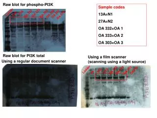
Strips 1 2 3 4 5 6 7 8 9 10 N M 1 2 3 4 5 6 7 8 9
RDB procedure • exons (or other regions of interest) • amplified by the polymerase chain reaction (PCR) • using labeled oligonucleotide primers • 5' biotin label on PCR primers
Amplicons • Amplification products • denatured • hybridized • with mutation specific DNA probes • covalently bound to solid membran
Incubation • nucleic acids: incubated with an enzyme conjugated to streptavidin. • enzyme-conjugated, streptavidin-biotin-nucleic acid complex is then washed • incubated with • a chromogenic • or luminogenic substrate, which allows visualization of hybridized spots
Materials and Methods • Total genomic DNA • extracted from peripheral blood leukocytes • Amniotic fluid cells (AF) • chorionic villi (CVS)
Oligonucleotide probes • A C6-amino-link phosphoramidite • amino moiety on the 5' end of the product
In vitro amplification of DNA by PCR Reaction mixture: • 5 µl template DNA • 5 µl forward primer (B-F27, 5 pmol/µl) • 5 µl reverse primer (R518, 5 pmol/µl) • 2.5 µl dNTP’s (2.5 mM of each dNTP) • 5 µl 10x PCR buffer • 1.5 µl 50 mM MgCl • 0.25 µl Taq polymerase • 23.75 µl water
PCR program: Our forward primer is biotinylated • 94°C for 5 min • 1 cycle • 94°C for 1 min • 50-55°C for 1 min • 72°C for 1 min • 30 cycles • 72°C for 5 min • 1 cycle • 4°C hold
Remarks • Repeated freeze thawing of the biotin labeled oligo or PCR products may damage the biotin label • Preferably the membrane should be stripped as soon as possible, but this can also be done a few days after the hybridization. • For chemiluminescent detection, the Solution A+B should be warmed to roomtemperature for at least 30 min
MATERIALS AND METHODS • PCR from 150 ng of genomic DNA • Preparation of membrane strips • Allele-specific hybridization and colordevelopment • Preparation of membrane strips
Preparation of membrane strips • Biodyne C (Pall Biomedical,U.S.A.) membrane • Membrane : activated briefly in 0·1 N HCl • Rinsedwith water and soaked in 16% 1-ethyl-3-[3-dimethylaminopropyl] • carbodiimide (EDC) for 15 min • it was rinsed in waterand air dried overnight • Oligonucleotide probes were dilutedwith 0·5 M NaHCO3/Na2CO3 buffer, pH 8·4(0.5 pmol/ml) for application onto the membrane.
Allele-specific hybridization and colour development • 50–60 ml of biotinylated-PCR product • Hybridizedwith the filter strips containing the normal and mutantprobes • in 0·8 ml hybridization buffer (2 ´ SSC,0·1% sodium dodecyl sulphate) (1 ´ SSC¼0·3 M NaCl,0·03 M sodium citrate)
Allele-specific hybridization and color development • sealed in a cooking pouch • Thepouch of reactants was denatured in boiling water for 5 min. • Hybridized at 428C ´ 1 h • Membrane strips were thenwashed in 0·4 ´ SSC,0·1% SDS at 428C for 10 min
Allele-specific hybridization and color development • The strips were then reacted at room temperature for 15 min with 20 ml streptavidin horse-radish peroxidase (Gibco BRL, as conjugate for the biotin-labelled hybridizationsignal) in 20 ml 2 ´ SSC, 0·1% SDS • washes(5 min ´ 2) in 2 ´ SSC, 0·1% SDS and (2 min ´ 2) in 0·1 Msodium citrate pH 5·0
Allele-specific hybridization and color development • Color development was carried out: • with 0·1% 3,30,5,50-tetramethylbenzidine dihydrochloride in • 0·1 M sodium citrate and 80 ml of 3% hydrogen peroxide for • 30 min at room temperature • The reaction was stopped : • rinsing once with 0·1 M sodium citrate and several timeswith water
Preparation of membrane strips • Approximately 4 ml was applied to each spot • allowed to dry for 15 min before fixation in 0·5 N NaOHfor 1 min • The membrane was then rinsed thoroughly with • water and air dried overnight • Membrane strips: stored at room temperature in adesiccator for up to 6 months.
Automated DNA sequencing Cd 2C>G
Haplotype analysis of the β-globin gene cluster from the patient's family.
PCR-RFLP 1 2 3 M 4 5 6 7
Direct genomic sequencing of the β-globin gene (ATG→AGG substitution of initiation codon)(a) The sequence of sense stranded sequence using Ex1 forward (b) The sequence of antisense stranded sequence using 3' reverse
Comparison of different factors determining the efficiency of ARMS and reverse hybridization in beta thalassemia diagnosis
Reference • J Clin Microbiol. 2001 March; 39(3): 871–878. Reverse Dot Blot Assay (Insertion Site Typing) for Precise Detection of Sites of IS6110 Insertion in the Mycobacterium tuberculosis Genome Lauren M. Steinlein and Jack T. Crawford* • Ian J Pub Heal. Spectrum of b-thalassemia Mutations in Isfahan Province of Iran (2007, in press) P Derakhshandeh-Peykar, H Hourfar, M Heidari, M Kheirollahi, M Miryounesi, and DD Farhud • Haemoglobin (2007, in press.) Distribution of ß-thalassemia mutations in Northern provinces of Iran. Derakhshandeh-Peykar P, Akhavan-Niaki H, Tamaddoni A, Ghawidel-Parsa S, Holakouie Naieni K, Rahmani M, Babrzadeh F, Dilmaghani-Zadeh M, Farhud DD (2007).
References • Lee GR, Forester J, Lukens J, Paraskovas F, Greer JP, Rodgers GM. The Wintrobe’s Clinic Hematology. Vol 1. 10th ed.Baltimore: Lippincott, Williams and Wilkins;1999. • Huisman THJ, Carver MFH. The beta- and delta-thalassemia repository. Hemoglobin. 1998; 22: 169-95. • Lorey FW, Arnopp J, Cunningham GC. Distribution of hemoglobinopathy variants by ethnicity in multiethnic • states. Genet Epidemiol. 1996; 13: 501-25. • Vetter B, Schwarz C, Kohne E, Kulozik AE. Beta- thalassemia in the immigrant and non-immigrant German populations. Br J Haematol. 1997; 97: 266-72.