Download

1 / 78
830 likes | 1.07k Views
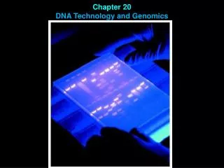
DNA Technology and Genomics. Why?. Understand the way in which plants and animals, including humans, develop, function and evolve Investigate the molecular basis of disease Develop products for medicine and crops for agriculture Solve crimes and paternity disputes

E N D
Why? • Understand the way in which plants and animals, including humans, develop, function and evolve • Investigate the molecular basis of disease • Develop products for medicine and crops for agriculture • Solve crimes and paternity disputes • Investigate endangered species for conservation management
Studying DNA • A number of methods have been developed that can be used to identify the DNA (genetic) profile of an individual • These methods can also be employed to measure genetic differences between individuals in a population • Techniques for working with DNA can be broken down into four major categories: • Copying DNA • Cutting and pasting DNA • Measuring DNA length • Probing DNA
Extracting DNA • Break open (lyse) the cells or virus containing the DNA of interest. This is often done by sonicating or bead beating the sample. Vortexing with phenol (sometimes heated) is often effective for breaking down protienacious cellular walls or viral capsids. The addition of a detergent such as SDS is often necessary to remove lipid membranes. • DNA associated proteins, as well as other cellular proteins, may be degraded with the addition of a protease. Precipitation of the protein is aided by the addition of a salt such as ammonium or sodium acetate. When the sample is vortexed with phenol-chloroform and centrifuged the proteins will remain in the organic phase and can be drawn off carefully. The DNA will be found at the interface between the two phases. • DNA is the precipitated by mixing with cold ethanol or isopropanol and then centrifuging. The DNA is insoluble in the alcohol and will come out of solution, and the alcohol serves as a wash to remove the salt previously added. • Wash the resultant DNA pellet with cold alcohol again and centrifuge for retrieval of the pellet. • After pouring the alcohol off the pellet and drying, the DNA can be re-suspended in a buffer such as Tris or TE.
Why we need so many copies • Biologists needed to find a way to read DNA codes. • How do you read base pairs that are angstroms in size? • It is not possible to directly look at it due to DNA’s small size. • Need to use chemical techniques to detect what you are looking for. • To read something so small, you need a lot of it, so that you can actually detect the chemistry. • Need a way to make many copies of the base pairs, and a method for reading the pairs.
Polymerase Chain Reaction (PCR) Used to massively replicate DNA sequences. Exploits enzymes and process of replication that normally occurs in cells. How it works: Separate the two strands with low heat Add some base pairs, primer sequences, and DNA Polymerase Creates double stranded DNA from a single strand. Primer sequences create a seed from which double stranded DNA grows. Now you have two copies. Repeat. Amount of DNA grows exponentially. 1→2→4→8→16→32→64→128→256… Polymerase Chain Reaction (PCR)
Polymerase Chain Reaction • Problem: Modern instrumentation cannot easily detect single molecules of DNA, making amplification a prerequisite for further analysis • Solution: PCR doubles the number of DNA fragments at every iteration 1… 2… 4… 8…
Polymerase Chain Reaction • Polymerase Chain Reaction (PCR) is broken down into three separate steps which are repeated until enough DNA is obtained (usually between 25 and 40 cycles) • Step 1 – Denaturation • Temperature is raised (94oC) in order to separate dsDNA into single strands • Step 2 – Annealing • Temperature decreased (50-60oC) in order for primers to anneal and provide a starting point for DNA polymerase • Step 3 – Extension • Temperature increased (72oC) which allows Taq polymerase to extend DNA • Taq polymerase is a heat resistant DNA polymerase isolated from the bacterium Thermus aquaticus which is found in hot springs and hydrothermal vents
Denaturation Raise temperature to 94oC to separate the duplex form of DNA into single strands
Design primers • To perform PCR, a 10-20bp sequence on either side of the sequence to be amplified must be known because DNA polymerase requires a primer to synthesize a new strand of DNA
Annealing • Anneal primers at 50-65oC
Annealing • Anneal primers at 50-65oC
Extension • Extend primers: raise temp to 72oC, allowing Taq pol to attach at each priming site and extend a new DNA strand
Extension • Extend primers: raise temp to 72oC, allowing Taq pol to attach at each priming site and extend a new DNA strand
Repeat • Repeat the Denature, Anneal, Extension steps at their respective temperatures…
RT-PCR: Variation of PCR • PCR reaction amplifies DNA from a single copy in the absence of cells • RT-PCR is a variant of PCR in which DNA is first reverse transcribed (copied in reverse) from RNA extracted from cells prior to being amplified by PCR as per usual protocol • Reverse transcription process involves the use of a reverse transcriptase enzyme and various other reagents including either a primer for a specific gene or an oligo-dT primer (string of Ts that will bind to poly-A tail of RNA. • Enzyme, reagent mix, primer and RNA are usually incubated at 37oC for 1 hour before the RT enzyme is inactivated at 72oC for approximately 15 minutes. The resulting cDNA is then used as the template for a PCR reaction.
PCR begins here RT-PCR
Vector DNA Cloning DNA to achieve copies • Use restriction enzymes and DNA ligase to insert the fragment of interest into the genome of another organism (e.g. bacteria) in order for it to multiply. • The resulting DNA is referred to as recombinant DNA as the genes from two different organisms are combined. • Once you have a large quantity of bacteria, you will be able to isolate a large quantity of the gene of interest
Restriction Enzymes • Discovered in the early 1970’s • Used as a defense mechanism by bacteria to break down the DNA of attacking viruses. • They cut the DNA into small fragments. • Can also be used to cut the DNA of organisms. • This allows the DNA sequence to be in a more manageable bite-size pieces. • It is then possible using standard purification techniques to single out certain fragments and duplicate them to macroscopic quantities.
Restriction Enzymes Definition: A restriction enzyme is a bacterial enzyme that recognises a short sequence of bases in a DNA molecule and cuts the DNA at this recognition site. • The position where a cutting enzyme can snip is its recognition sequence and is where a particular order of nucleotides occurs. • Some restriction enzymes cut the two strands of a DNA molecule at points directly opposite each other to produce cut ends that are ‘blunt’. • Other cutting enzymes cut one strand at one point, but cut the second strand at a point that is not directly opposite. • The overhanging cut ends made by these cutting enzymes are called ‘sticky’. These sticky ends are complementary.
Pasting DNA • Once separated, DNA fragments from different sources can be joined (ligated) together. • Sticky ended fragments will initially join by hybridization or complementary base pairing. • Bonds within the single strands of DNA are then repaired by DNA ligase (this is similar to the action of DNA ligase in linking of Ozaki fragments on the lagging strand during DNA replication)
Gel Electrophoresis • A copolymer of mannose and galactose, agaraose, when melted and recooled, forms a gel with pores sizes dependent upon the concentration of agarose. • The phosphate backbone of DNA is highly negatively charged, therefore DNA will migrate in an electric field.
Gel Electrophoresis • The size of DNA fragments can then be determined by comparing their migration in the gel to known size standards • Ethidium bromide or other dyes that bind to DNA are added prior to electrophoresis in order to visualize migration of DNA • Ethidium bromide flouresces bright orange when exposed to UV light
Reading DNA – DNA Sequencing • DNA sequencing reactions are just like the PCR reactions for replicating DNA with the exception that the reactions are run in the presence of a dideoxynucleotide. • Dideoxyribonucleotides are the same as nucleotides, with one exception. They do not have 3' hydroxyl group, so once a dideoxynucleotide is added to the end of a DNA strand, there's no way to continue elongating it. • Sequencing reactions are set up in groups of four, e.g. one containing dideoxy-A, one containing dideoxy-C, one containing dideoxy-G and one containing dideoxy-T. Each reaction tube contains a mix of normal nucleotides (A,C,G, T) and a small amount of the particular dideoxynucleotide. • Taking dideoxy-C as an example, replication of DNA will occur as per a PCR reaction. MOST of the time when a ‘C' is required to make the new strand, the enzyme will get a good one and there's no problem. MOST of the time after adding a C, the enzyme will go ahead and add more nucleotides. However, 5% of the time, the enzyme will get a dideoxy-C, and that strand can never again be elongated. It eventually breaks away from the enzyme, a dead end product.
Reading DNA – DNA Sequencing • Sooner or later ALL of the copies will get terminated by a T, but each time the enzyme makes a new strand, the place it gets stopped will be random. In millions of starts, there will be strands stopping at every possible T along the way. • ALL of the strands we make started at one exact position. ALL of them end with a T. There are billions of them ... many millions at each possible T position. To find out where all the T's are in our newly synthesized strand, all we have to do is find out the sizes of all the terminated products!
Reading DNA – DNA Sequencing • Gel electrophoresis can be used to separate the fragments by size and measure them. • The dideoxynucleotides present in the fragments have been labelled with a radioisotope or a flourescent dye. In the case of the latter, these can be read by a laser and the information feed back to computer. • Following electrophoresis and visualization of fragments we can determine the sequence. Smallest fragments are at the bottom, largest at the top. The positions and spacing shows the relative sizes. At the bottom are the smallest fragments that have been terminated by dideoxynucleotides.
Assembling Genomes • Based on sequencing data, we can take fragments and put them back together. Not as easy as it sounds!!!!! • SCS Problem (Shortest Common Superstring) • Some of the fragments will overlap • We try to fit overlapping sequences together to get the shortest possible sequence that includes all fragment sequences • Problems that may arise during this process: • DNA fragments contain sequencing errors • There are two complements of DNA – we need to take into account both directions of DNA • The repeat problem - 50% of human DNA is just repeats. If you have repeating DNA, how do you know where it goes?
Analyzing a Genome • How to analyze a genome in four easy steps. • Cut it • Use enzymes to cut the DNA in to small fragments. • Copy it • Copy it many times to make it easier to see and detect. • Read it • Use special chemical techniques to read the small fragments. • Assemble it • Take all the fragments and put them back together. This is hard!!! • Bioinformatics takes over • What can we learn from the sequenced DNA. • Compare interspecies and intraspecies.
Nucleotide Hybridization • Single-stranded DNA or RNA will naturally bind to complementary strands. • Hybridization is used to locate genes, regulate gene expression, and determine the degree of similarity between DNA from different sources. • Hybridization is also referred to as annealing or renaturation. • Hybridization uses oligonucleotides to find complementary DNA or RNA seqments. Oligonucleotides are single-stranded DNA molecules of 20-30 nucleotides in length. • Oligonucleotides are made with DNA synthesizers and tagged with a radioactive isotope or fluorescent dye • Molecular techniques based on hybridization include Southern blotting, Northern blotting and microarrays.
Create a Hybridization Reaction 1.Hybridization is binding two genetic sequences. The binding occurs because of the hydrogen bonds [pink] between base pairs. 2. When using hybridization, DNA must first be denatured, usually by using use heat or chemical. T C T A G C G T C A T T G T TAGGC ATCCGACAATGACGCC
Create a Hybridization Reaction Cont. 3. Once DNA has been denatured, a single-stranded radioactive probe [light blue] can be used to see if the denatured DNA contains a sequence complementary to probe. 4. Sequences of varying homology stick to the DNA even if the fit is poor. ACTGC ACTGC ATCCGACAATGACGCC Great Homology ACTGC ATCCGACAATGACGCC ATTCC Less Homology ATCCGACAATGACGCC ACCCC Low Homology ATCCGACAATGACGCC
Southern Blotting • Cut total genomic DNA with restriction enzymes and separate by electrophoresis • Blot the fragments onto nitrocellulose filter paper (the Southern blot) • Probe the blot for a particular DNA region of interest using a specific labelled oligonucleotide. • Wash blot to remove oligonucleotide that has not bound. • Identify gene or region of interest by visualizing regions where probe has hybridised with DNA on Southern blot. • Northern blotting follows a similar process to Southern blotting except that RNA is run on the initial gel and the oligonucleotide probes are used to detect expression of particular genes.
DNA Microarray Affymetrix Microarray is a tool for analyzing gene expression that consists of a glass slide. Each blue spot indicates the location of a PCR product. On a real microarray, each spot is about 100um in diameter.
DNA Microarray Tagged probes become hybridized to the DNA chip’s microarray. Millions of DNA strands build up on each location.
DNA Microarrays • An array works by exploiting the ability of a given mRNA molecule to hybridize to the DNA template. • Using an array containing many DNA samples in an experiment, the expression levels of hundreds or thousands genes within a cell by measuring the amount of mRNA bound to each site on the array. • With the aid of a computer, the amount of mRNA bound to the spots on the microarray is precisely measured, generating a profile of gene expression in the cell.
An experiment on a microarray In this schematic:GREENrepresents Control DNA REDrepresents Sample DNA YELLOWrepresentsa combination of Control and Sample DNA BLACKrepresents areas whereneither the Control nor Sample DNA Each color in an array represents either healthy (control) or diseased (sample) tissue. The location and intensity of a color tell us whether the gene, or mutation, is present in the control and/or sample DNA. 10
Forensics:DNA technology in action • Each of us is genetically unique, with the exception of identical (monozygotic) siblings. While phenotypic differences are apparent among us, the most fundamental expression of our uniqueness is in our genetic material, DNA. • Today, individuals can be identified through a technique known as DNA profiling. • The amount of DNA needed for DNA profiling is very small because DNA can be amplified through the polymerase chain reaction (PCR). • One person’s DNA profile is constant, regardless of the type of cell used to prepare the profile. A DNA profile prepared from a person’s white blood cells is identical to that prepared from the same person’s skin cells or other somatic cells. • Because DNA molecules are only slowly degraded, DNA profiling can be carried out on biological samples from crime scenes from years ago, and this profiling has led to the solution of many ‘cold cases’ worldwide.
Forensic Identification • Identification using DNA is a powerful tool that can be applied in many situations including: • forensic applications • Can the DNA found at a crime scene be matched to a person on the national DNA database? • Is this blood spot from the victim or from the possible assailant? • In a rape case, is this semen from a previously convicted rapist? • mass disasters, such as passenger aircraft crashes, the 9/11 terrorist attacks, the Bali bombings • Can the various remains that have been recovered be matched to a particular person known to have been on-site? • identification of human remains • Are these remains those of a particular missing person? • Who was the unknown child, tagged as body number 4, recovered after the sinking of the Titanic in 1912?
What DNA is used for identification • Depending on the purpose and circumstances of the identification, the DNA used comes from either the chromosomes (nuclear DNA) or from mitochondria (mtDNA). • In both cases the identification depends on the existence of segments of DNA that vary greatly between individuals. Such regions of DNA are termed hypervariable. • Hypervariable regions of DNA that are currently used for identification are: • short tandem repeats (STRs) in the nuclear DNA, also known as microsatellites. • hypervariable regions (HVRs) in the non-coding region of mtDNA.
STRs and HVRs Short Tandem Repeats (STRs) in the nuclear DNA, also known as microsatellites. • A large number of STRs are present on different human chromosomes. • DNA from STRs can identify one person uniquely (apart from identical siblings). • DNA samples from relatives are not required. • Used when there is a need to match a DNA sample from a crime scene to just one particular person. Hypervariable Regions (HVRs) in the non-coding region of mtDNA. • mtDNA identification is less precise because persons from the same maternal line have identical mtDNA profiles. • mtDNA is used only when chromosomal DNA cannot be recovered or when chromosomal DNA is degraded because of age. • Identification using mtDNA is mainly applied either • to identify victims of mass disasters where the names of the victims are known but where identification of the remains by conventional means, such as visual inspection or dental records, cannot be done, or • to identify decomposed remains when the identity is suspected to be one of a few particular missing persons. In both cases, there must be living relatives on the maternal line to provide mtDNA for comparison with the mtDNA from the remains.
DNA Fingerprinting - HVRs • The original technique of identification using DNA was called DNA fingerprinting and was developed as an identification tool in 1985 by Professor Sir Alec Jeffreys • This technique used DNA from hypervariable regions, known as minisatellites, that are located near the ends of chromosomes. Minisatellites are chromosomal regions where sequences of 9 to 80 base pairs are repeated tens or hundreds of times. • DNA fingerprinting involved cutting minisatellites from the chromosomal DNA with a restriction enzyme (Hin fI), separating the DNA fragments by electrophoresis, transferring them to a membrane using Southern blotting and exposing the fragments to one probe that hybridised to a base sequence present in all the minisatellites. • This probe, known as a multi-locus probe, carried a radioactive label. The final result seen on an autoradiograph was a pattern of up to 36 bands, something like a barcode, with each band being one allele of one of the minisatellites. Because of the variation between individuals, each DNA fingerprint is unique. The figure above shows the simplified DNA fingerprints of two people based on just four hypothetical minisatellites. In actual DNA fingerprinting, the pattern for each individual has many more bands.
DNA profiling - STRs • DNA fingerprinting has been replaced by a technique known as DNA profiling that uses short tandem repeats (STRs). These are • STRs are hypervariable regions of chromosomes where sequences of just two to five base pairs are repeated over and over. These regions are very common and hundreds are scattered throughout the human chromosomes. • STRs are termed ‘short’ because the repeat sequences are only 2 to 5 base pairs long, and ‘tandem’ because the repeats occur one after the other. However, the number of repeats at an STR locus can vary between people and each variation is a distinct allele. • The number of repeats of a 4-base pair sequence at one STR locus on the number-5 chromosome ranges from 7 to 15. • In most cases, the alleles at an STR locus on a human chromosome are named according to the number of repeats and so are identified as allele 7, allele 8 and so on. The figure below shows an STR with 7 repeats of the sequence CATT.
DNA profiling - STRs • At each STR locus, one individual is either homozygous or heterozygous and so can have a maximum of just two different alleles. These alleles are inherited in a Mendelian fashion. • The figure below shows that a person who is heterozygous 5/7 at one particular STR locus has one allele with 5 repeats and another allele with 7 repeats. • Within the gene pool of a population, however, many different alleles can exist at each STR locus.
Frequencies of the alleles at the D5 STR locus on the number-5 chromosome for three sample populationsin Australia. Note that the allele frequencies vary within a population, with allele 11 being far more common than allele 15. Note also that the frequencies vary between populations, with allele 7 being about 20 times more common in Asian populations than in the other two populations.
Why use STRs rather than minisatellites? • Compared with DNA fingerprinting, DNA profiling: • is far more sensitive and requires smaller quantities of DNA (even a pinhead sized spot of blood can provide sufficient DNA) and the STRs can be amplified by the polymerase chain reaction (PCR) • is based on alleles whose sizes allow fragments differing by just one base pair to be distinguished • is carried out in a much shorter time — hours rather than days • uses several single-locus probes rather than one multi-locus probe • uses coloured fluorescent labels to visualise the STRs rather than radioactive labels so that each different STR allele can be identified by colour as well as by size • produces less complex patterns that are more easily interpreted • In addition, unlike minisatellites, population data on allele frequencies of STR alleles can be obtained.
DNA Profiling in Australia • All Australian states use a common method of DNA profiling for forensic purposes that involves nine STRs from different human chromosomes. • These STR markers were chosen for this purpose because they are reproducible and robust, easy to score, are highly informative and have low mutation rates. • In addition, a tenth marker (that is not an STR) is used to identify the gender of the individual. This gender marker is the Amel locus that is present on both the X chromosome and the Y chromosome. • The Amel gene on the X chromosome is just 107 base pairs long while that on the Y chromosome contains 113 base pairs. As a result, the gender of a person can be identified from this marker.
Loci currently used for DNA profiling in Australia For simplicity, STR loci that start with the letter D are identified by their chromosomal location only, for example, D13 or D7. In reality, the naming of STRs is more complex because there are multiple STR regions on the one chromosome and these two STRs (D13 and D7) are formally identified as D13S317 and D7S820.