Download
1 / 61
610 likes | 793 Views
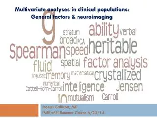
Multivariate analyses in clinical populations: General factors & neuroimaging. Joseph Callicott, MD fMRI/MRI Summer Course 6/20/14. Introduction. The ‘Age of B ig Data’

E N D
Multivariate analyses in clinical populations: General factors & neuroimaging Joseph Callicott, MD fMRI/MRI Summer Course 6/20/14
Introduction The ‘Age of Big Data’ Lohr, “GOOD with numbers? Fascinated by data? The sound you hear is opportunity knocking…” (NY Times, 2/22/2012) We routinely collect ‘multimodal data’ E.g., mood rating scale and structural MRI Compile or compare, but typically without multimodal analysis Projects classified as ‘geno-,’ ‘proteo-,’ or ‘pheno-’ already connotes ‘big data:’ Each fMRI image presents ~20K analyses GWAS model = strict correction for multiple comparisons Current model = parallel correlation/association per dataset Proposed model = multivariate approach w/data reduction Simplified analyses Smaller statistical ‘cost’ Some current theoretical approaches become testable’ (RDoC)
Outline A Tale of Two Lectures: Imaging genetics & schizophrenia Relevant issues for clinical populations Why imaging genetics? Imaging genetics 101 Multivariate analyses of fMRI: within experimental dataset General vs specific factors in data g “i” : factor analytic solution of general factors in fMRI task data Multivariate analyses of fMRI redux: across experimental dataset
Issues of special interest to clinical studies… BOLD fMRI in clinical populations: BOLD fMRI is not, strictly speaking, a clinically informative measure No pathognomic findings, to date Performance likely to differ fundamentally in tasks that HVs will perform near ceiling I.e., ~100% accuracy and faster RT than patients BOLD fMRI in healthy subjects ‘predictive relationships’ between behavior and BOLD implicit in design, perhaps not strongly correlated with group map activation Genetic associations to BOLD (the bulk of the imaging genetics literature) do not necessarily connote a ‘real’ effect of a given polymorphism Is the phenotype heritable? In past, twin or sibling studies Currently, within ‘only’ HV = GCTA (Visscher) (Callicott et. al. 2000; Manoach et al., 2000; Callicott et al. 2003; others) (Van Snellenberg et al. 2006)
So, then, why imaging genetics? • Crass commercial message: • Simple plan, high impact • Have an fMRI task in a relatively large sample? • Healthy controls preferable • N > 40 • Draw blood or swab cheek • Genotyping at most resolutions fast & cheap* • In SPM: ANCOVA or regression suffice and seem reasonably powered COMT led from primate to human imaging, and then to drugs targeting cognitive impairments
Seriously, though, why imaging genetics? • Few routes to neural mechanism using in vivo human data • Animal model • Drug study • BOLD fMRI (MRSI, MEG, EEG) • Genes do not code for mental illness, per se • Genes code for heritable aspects of brain function, intermediate- or endo-phenotypes • Genetic risk for illnesses like schizophrenia is polygenic, heterogeneous • Gene interact with each other and the environment • BOLD fMRI, as an alternate metric of specific or general cognitive systems, offers ‘real world validation’ • Putative genetic mutations (including private mutations (CNVs) • In spite of growing sample sizes, association studies risk false positives • RDoC domains and constructs • If these do not correspond to brain systems we can map, then may be as doomed as DSM
Take home message… Larger samples needed (GWAS), typically via collaboration across centers (ENIGMA) BIG DATA is here Multivariate or non-hypothesis-driven analyses offer the potential for novel, highly informative findings CNV & cognition Very good software often freely available: PLINK AFNI GingerALE-SLEUTH-MANGO R (many) (Stefansson et al., Nature, 2014)
(*Visscher et al., 2010; Nan et al. 2012**; Postuma et al. 2002***; McGue& Bouchard, 1998#;^Burmeister et al., 2008)
Interest in imaging genetics predicated on heritability of phenotypes… Patients > Controls (N=13) (N=18) Callicott et al. Cereb Cortex 2000 Healthy Siblings > Controls (N=48) (N=33) PFC BOLD during our Nback h2 = 0.4-0.5 Callicott et al.Am J Psychiatry 2003 Blokland et al.Biol Psych 2009, J Neurosci 2011; Koten et al. Science 2009
Finding genes for highly heritable, but complex diseases • Remains difficult, even when n=100K • Caused by many (100-1000s) of genes • The effects of a mutation vary between • people affected person Has all the genes (note this doesn’t mean the exact same set) unaffected May still carry some genes (like a parent of a sick person) “nonpenetrant” Has all of the genes but is NOT sick for reasons we can’t explain (Goldman et al. Nat Rev Genet 2006)
Catechol O-methyltransferase (COMT): NIMH Intramural Success Story (Meyer-Lindenberg et al. 2006) (Egan et al. 2001) (Apud et al. 2006)
Functional impact COMT Val105/158Met val/met rs4680 5’ • Now validated at multiple levels: • Animal models: • Reduced enzymatic activity • Altered synaptic dopamine levels • Human data: • Reduced enzymatic activity in vitro lymphoblastoid cell lines • Altered transcription/reduced activity post mortem • Altered D1 but not D2 receptor density in PFC • PFC efficiency in BOLD fMRI • Combined: • Sex effects mostly in males ((Papaleo et al., 2014) striatum mammalian PFC
Physiological efficiency Executive cognition genotype effect F=5.41, df= 2, 449; p<.005. vv>vm>mm, SPM 99, p<.005 Effect of rs4680 on frontal lobe function Replicated but n’s ~20 n = 181 n = 218 n = 58 (Egan et al PNAS2001) Circa 2014: How have these findings held up?
BOLD phenotypes in simple association: COMT and PFC (Mier, Kirsch, Meyer-Lindenberg; 2009)
BOLD phenotypes in simple association: 5-HTTP and Amygdala (Munafo, Brown, and Hariri; 2007))
BOLD phenotypes in simple association: Power? (Barnett et al, 2008)
1st generation imaging genetics: simple association • Candidate genes • KIBRA impaired memory & expressed in hippocampus (Papassotiropoulos et al., Science 2006) • Replication in 3 independent populations in behavioral memory measures • In 30 healthy subjects, KIBRA associated with reduced hippo activation
2ndgeneration imaging genetics: GWAS era • Genetic mutations modeled in cell culture or animals • Association based on disease GWAS (ZNF804A) • Esslinger et al. 2009 (Science) • Rasetti et al. (Arch Gen Psychiatry)
2ndgeneration imaging genetics: Epistasis and pathways PFC neuronal function: ‘optimized’ by dopamine & GABA interactions (Goldman-Rakic & Selemon 1997) (Seamans et al., 2001)
2ndgeneration imaging genetics: Epistasis (Straub et al., 2007)
2ndgeneration imaging genetics: Translational neuroscience • Epistasis (gene-gene interaction) • Initially based on candidate-by-candidate • Buckholtz et al., Mol Psychiatry 2007 • Data-driven (machine learning) • Nicodemus et al., Hum Genet 2010 • Now predicated on detailed cellular or animal modeling • COMT x DTNBP1 (Papaleo et al., 2013) • DISC1 x NKCC1 (Kim et al., Cell 2012 & Callicott et al. J Clin Invest 2013) COMT x Dysbindin interaction BOLD fMRI Left DLPFC (a.u.) COMT: V/V V/M M/M V/V V/M M/M V/V V/M M/M Bray Hap: -/- -/- -/- +/- +/- +/- +/+ +/+ +/+
Next generation imaging genetics? More of the same (‘sophisticated univariate’)? Network/connectivity? Hypothesis-free? Novel phenotypes (processing speed) ICA/PCA/CPCA networks Hypothesis-free pattern detection (random forest)
Sophisticated univariate: Imaging GWAS • BOLD fMRI GWAS • Nback (n = 364) • Illumina 650K chip genotyping • Automated extraction of AAL ROIs • First GWAS + using BOLD fMRI (Callicott, Spencer, et al., in prep)
Sophisticated univariate: Novel questions As a heritable trait, BOLD fMRI phenotypes show other sensitivities….. • Long history within animal literature showing significant effects of environment on brain structure & function • Beneficial effects of ‘enrichment’ (toys, limit isolation) (Hebb, Am J Psychiatry, 1955) • fMRI during social stress task influenced by environment • Urban upbringing or urbanicity linked to increased risk for mental illness (Van Os et al., Nature, 2010) (Lederbogenet al., Nature, 2012)
Sophisticated univariate: Imaging G x E As a heritable trait, BOLD fMRI phenotypes show other sensitivities….. • fMRI during WM ( 3 cohorts (USA1 = 124; USA2 =92; Italy1=226 ) • Sensitivity to childhood environment (Urbanicity) (Ihne et al., in submission)
Sophisticated univariate: Imaging G x E • fMRI during WM ( 3 cohorts (USA1 = 124; USA2 =112; Italy1=226 ) • Gene-by-environment interaction (COMT x Urbanicity) (Ihne et al., in submission) Ihne et al., in preparation
Constrained principle component analysis (CPCA) (David AA Baranger– Wash U) Multivariate network analysis: ICA/PCA/CPCA banish ‘blob-ology’ • Todd Woodward and colleagues, University of British Columbia: • CPCA provides a “unified framework [for]… regression analysis and principal component analysis .” • To identify functional systems using from singular-value decomposition of BOLD time series, • These systems are imaged by constraining analyzed BOLD signal from a particular interval of time against all other scans (i.e., all others are baseline) http://www.nitrc.org/projects/fmricpca
CPCA • Z or ‘activation’ matrix = individual time series for all subjects (rows) for all voxels in the brain (columns) • Our standard SPM5 via XNAT first level processing of 0B alternating with 2B • G or ‘design’ matrix = a model to predict BOLD signal changes (columns) over all fMRI scans (rows) • SPM5 often uses a canonical hemodynamic response function (HRF) to deconvolve signal, fMRI-CPCA uses finite impulse response function (FIR) http://www.nitrc.org/projects/fmricpca
CPCA • N-back model not complicated: • Simply provide onset and offset of 0B and 2B task epochs • Components = extracted components represent networks • Component loadings= loosely, correlation coefficients between component scores and BOLD signal that was predicted from imposed constraints (design) http://www.nitrc.org/projects/fmricpca
CPCA • Identify and then display components using MRICon for anatomical localization (http://www.nitrc.org/projects/mricron) • In this case, not really using estimated hemodynamics • Rather, we wish to compare effect of diagnosis or genotype using component scores and predictor weights • Predictor weight = contribution of G matrix to changes in components over the fMRI time series (~ correlation of component score and g)
CPCA: Confusing Problematic Conflicting Agonizing • Unspecified error required recalculation of component weights • Same networks found with addition of a fourth DMN • Differentiates NC and SIBs from SCZ • No longer appears to be identifying intermediate phenotype
CPCA: Nback systems Anti-task Network resembles cingulate from DMN + hippocampus WM Network DLPFC +parietal Motor system Anti-task #2 resembles parietal From DMN + cerebellum
CPCA: Factors sensitive to disease, not genetics • Unspecified error required recalculation of component weights • Same networks found with addition of a fourth DMN • Differentiates NC and SIBs from SCZ • No longer appeared to identify intermediate phenotype p<0.05 p<0.05
CPCA: Not particularly sensitive in general • 420 HV • CPCA (2back) = 4 factors • Neuro- = 6 cognitive factors • 2B as measured in lab • g estimates
Big data benefits reproducibility… ENIGMA: first GWAS+ sMRI (Stein et al. 2009, 2010; Thompson et al., 2014) Heritability for novel phenes Replication on large scale
Outline A Tale of Two Lectures: Imaging genetics & schizophrenia Why imaging genetics? Imaging genetics 101 Multivariate analyses of fMRI: within experimental dataset General vs specific factors in data g “i” : factor analytic solution of general factors in fMRI task data Multivariate analyses of fMRI redux: across experimental dataset
The general cognitive factor (Spearman’s g) (Jensen, 1998) (Dickinson et al., 2008)
Where is g? Various conceptual, functional and structural support for PFC and PAR (at minimum) Barbey et al. (Brain 2012)found similar results in 182 focal brain lesion patients ‘Lesion maps’ from 241 patients w/ focal brain damage and g (Gläscher et al. PNAS 2010).
Is g associated with fMRI activation? Nback(n= 161) higher g greater efficiency Replication (n= 582) higher g greater efficiency Exact overlap Notes: Analysis: SPM5 multiple regression controlling for age, sex 2B accuracy g (r = 0.3, p < 0.001)
Replication…cont’d Areas within replication exactly overlapping discovery… discovery Replication 3 Replication 4 Replication 5 Replication 4 (n= 393) Replication 5 (n= 306) Replication 3 (n= 211)
But… DSVTv g Faces v g MTL v g
g correlates with similar areas across 4 tasks in same 161 HVs Notes: Analysis: SPM5 multiple regression controlling for age, sex 161 with QC+ NB, MTL, Faces, DSVT NB as discovery ROI, others queried at p < 0.05 Nback(n= 161)
Is there a general solution for fMRI? • 161 HVs with QC+ Nback, MTL (incidental encoding), Faces (response to aversive faces), and DSVT (processing speed) • Individual 1st level maps created for each task • Sue Tong: automated script to extract parameter estimates in Automated Anatomical Labeling (AAL) ROIs • Mean fMRI ‘signal” transformed to Z score • Factor analysis: • Principle component extraction • Orthogonal and oblique rotations • Factor scores estimated • i (fMRI g) = sum of factor scores • Comparison across task and against cognitive measures (big g)
fMRI (Nback) i (161 HVs, max likelihood extraction w/ varimax rotation, 60.1% total variance explained, goodness-of-fit p < 1e-5) .30 .50 .41 .42 .26 .40 .42 .16 .13 .10 .11 .13
fMRI (Nback) i .54 .79 .59 .90 .43 .7 .27 .36 .56 .52 (161 HVs, max likelihood extraction w/ varimax rotation, 60.1% total variance explained, goodness-of-fit p < 1e-5)