Management of HIV-Positive Patient with Gastric B-Cell Lymphoma and Anemia
A 48-year-old Black male with a history of HIV presents with left-sided abdominal pain, fever, and fatigue. His examination reveals enlarged inguinal nodes, splenomegaly, and scattered Kaposi's sarcoma lesions. Laboratory findings indicate severe immunosuppression, and a CT scan reveals a gastric mass and enlarged lymph nodes. A biopsy confirms large B-cell non-Hodgkin's lymphoma. Despite chemotherapy treatment, he develops anemia. Ongoing management includes supportive care and potential retreatment strategies to address recurrent lymphoma.


Management of HIV-Positive Patient with Gastric B-Cell Lymphoma and Anemia
E N D
Presentation Transcript
Patient Description • 48 year old, black male with history of HIV and Kaposi’s sarcoma presents with left sided abdominal pain, fever and fatigue worsening over past month • On lamivudine/zidovudine, efavirenz, TMP-SMX, azithromycin, but on no KS therapy
Patient Description • Enlarged inguinal nodes bilaterally, palpable spleen, diffuse abdominal pain with some guarding, no palpable masses, active BS, no rectal masses and hemoccult negative. • Few scattered KS lesion on lower extremities bilaterally, no edema • Remainder of exam normal
Patient Description • CD4+ count 56 cells/mm3; plasma HIV RNA 1000 copies/mL • Liver enzymes mildly elevated, creatinine normal, amylase normal • Hb 9.7 g/dL; Hct 29.3%; WBC 3000/mm3; platelet count 120,000; MCV 90 fL; retic 0.014
Possible Diagnosis 1. GI Kaposi’s sarcoma with GI bleed 2. Non-Hodgkin’s lymphoma 3. Abdominal abscess, MAI or others 4. CMV colitis 5. Anal cancer 6. Progressive HIV/AIDS
Possible Diagnosis 1. GI Kaposi’s sarcoma with GI bleed 2. Non-Hodgkin’s lymphoma 3. Abdominal abscess, MAI or others 4. CMV colitis 5. Anal cancer 6. Progressive HIV/AIDS
Additional Tests 1. CT abdomen 2. Biopsy lymph node 3. Repeat VL and HIV genotype 4. Culture blood and stool, endoscopy 5. Stool occult blood, bone marrow, further heme work up 6. Chest X-ray and Chest CT if appropriate
Additional Tests 1. CT abdomen 2. Biopsy lymph node 3. Repeat VL and HIV genotype 4. Culture blood and stool, endoscopy 5. Stool occult blood, bone marrow, further heme work up 6. Chest X-ray and Chest CT if appropriate
Laboratory Findings • Urinalysis normal, bilirubin normal, urine hemosiderin negative, LDH 500 IU/L, G6PD level normal, ferritin level elevated. • Stool occult blood positive, blood cultures X 3 neg, MAC cultures neg, AFB negative, stool cultures neg, CXR normal • CT scan abdomen - ordered • HIV drug genotype - ordered • Bone marrow aspiration and biopsy- ordered
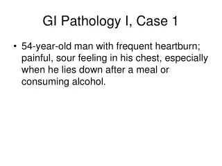
Laboratory Findings • CT scan of abdomen shows large gastric mass, enlarged perigastric and periaortic nodes, a few liver nodules and small amount of ascites • Gastroscopy shows large gastric ulcerated mass, no active bleeding • Histology reveals large B-cell NHL, CD-20+, no CMV or KS
Gastric lymphoma. Persistent gastric wall thickening (S). Enlarged spleen and an enlarged Lt. gastric lymph node (arrow)
Clinical Decision Point • The patient has no CNS symptoms, CBC has not changed. Staging work up includes: • CT of chest - no abnormalities • LP with CSF cytology - neg for lymphoma • Bone marrow aspiration and biopsy • hypocellular, normal iron stores, no lymphoid infiltrates, normal lymphocyte flow panel, no granuloma or infection seen
Clinical Decision • Chemotherapy (e.g CHOP-R or EPOCH-R) • Hydrate, allopurinol, follow electrolytes, creatinine, phosphorus, calcium • Intrathecal cytosine arabinoside X 4 • Continue TMP-SMX, azithromycin • Begin prophylaxis with ciprofloxacin • Follow CBC, +/- G-CSF, +/- rEPO • Change ART • All of the above
Clinical Decision • Chemotherapy (e.g CHOP-R or EPOCH-R) • Hydrate, allopurinol, follow electrolytes, creatinine, phosphorus, calcium • Intrathecal cytosine arabinoside X 4 • Continue TMP-SMX, azithromycin • Begin prophylaxis with ciprofloxacin • Follow CBC, +/- G-CSF, +/- rEPO • Change ART • All of the above
Clinical Course • Patient tolerates chemotherapy with EPOCH-R, however after two cycles, Hb now 8.0 g/dL. What do you do? 1. Repeat bone marrow aspirate and biopsy 2. Give iron and folic acid 3. Check EPO level and give recombinant erythropoietin alpha 4. Stop AZT and switch to new ARV 5. Transfuse 2 units of PRBC and schedule endoscopy
Clinical Course • Patient tolerates chemotherapy with EPOCH-R, however after two cycles, Hb now 8.0 g/dL. What do you do? 1. Repeat bone marrow aspirate and biopsy 2. Give iron and folic acid 3. Check EPO level and give recombinant erythropoietin alpha 4. Stop AZT and switch to new ARV 5. Transfuse 2 units of PRBC and schedule endoscopy
Clinical Course • EPO level 125, rEPO administered at 40,000 IU per week with supplemental iron and folic acid. • After three cycles of EPOCH, patient has achieved a complete remission. Treatment continues for 6 cycles. CBC returns to normal.
Clinical Course • 6 months later, he is again noted to have Hb 9.5 g/dL, WBC 2300/mm3 and platelets 65,000/mm3 and enlarged femoral nodes • What do you do?
What do you do? • Work up anemia, DC TMP-SMZ, retreat with epoetin alfa and follow nodes • Assume recurrent lymphoma and retreat with EPOCH-R • Assume lymphoma and treat with alternate regimen • Assume progressive KS and treat with liposomal doxorubicin • Biopsy lymph node and bone marrow
What do you do? • Work up anemia, DC TMP-SMZ, retreat with epoetin alfa and follow nodes • Assume recurrent lymphoma and retreat with EPOCH-R • Assume lymphoma and treat with alternate regimen • Assume progressive KS and treat with liposomal doxorubicin • Biopsy lymph node and bone marrow
Clinical Course • Biopsy of lymph node and bone marrow shows high grade B-cell (CD20+) NHL, large cell type • Patient treated with ESHAP, G-CSF, epoetin alfa and prophylactic antibiotics (ciprofloxacin) • Complete remission achieved after 5 cycles, patient treated for 8 cycles and continues to be followed.
Key Points • AIDS patients can have multiple cancers • Evaluate for multiple etiologies for anemia in advanced AIDS patients • Consider use of Epoetin alfa when risk of further myelosuppression is great • Early relapse in AIDS/NHL should be treated with non-cross resistant salvage chemotherapy • Use prophylactic antibiotics and hematopoietic growth factors in AIDS patients on chemotherapy, especially if receiving Rituximab
Patient Description • 57 year-old white male with HIV infection since 1985, currently on tenofovir/emtricitabline,darunavir/ritonavir,TMP-SMX, azithromycin, valgancyclovir and fluconazole • CD4 count 12, VL >150,000 copies/mL, Hb 9.2 g/dL, Hct 28 %, platelet count 111,000/mm3 • Presents with low grade fevers, progressively worsening personality changes for past month and mental confusion and lethargy for past week
Patient Description • He suffers grand mal seizure on day of admission • MRI scan of brain shows single contrast-enhancing lesion in the basal ganglion with surrounding edema • What do you suspect is the most likely diagnosis?
Possible Diagnosis 1. CMV encephalitis 2. Toxoplasmosis 3. PML 4. CNS lymphoma 5. Fungal abscess 6. Infectious meningitis
Possible Diagnosis 1. CMV encephalitis 2. Toxoplasmosis 3. PML 4. CNS lymphoma 5. Fungal abscess 6. Infectious meningitis
Clinical Decision Point • Patient loaded with Phenitoin and started on dexamethasone 10 mg QID • Which diagnostics study would produce the greatest likelihood of a diagnosis?
Clinical Decision Point Which diagnostics study would produce the greatest likelihood of a diagnosis? 1. Toxoplasma serology 2. Brain biopsy 3. LP with CSF cultures and cytology 4. LP with toxo titer, CMV and EBV PCR 5. Blood cultures for bacteria, fungi, AFB, viruses 6. Bone marrow aspiration and biopsy 7. None of the above
Clinical Decision Point Which diagnostics study would produce the greatest likelihood of a diagnosis? 1. Toxoplasma serology 2. Brain biopsy 3. LP with CSF cultures and cytology 4. LP with toxo titer, CMV and EBV PCR 5. Blood cultures for bacteria, fungi, AFB, viruses 6. Bone marrow aspiration and biopsy 7. None of the above
Clinical Course • CSF cultures and cytologies negative; CMV and EBV CSF PCR sent • Blood cultures sent, preliminary negative • Toxo IgG+ but IgM negative • Sterotactic biopsy under MRI guidance shows immunoblastic lymphoma, EBV+, CD20+, HHV-8 negative • Retic count 0.14, LDH 140 IU/L, ferritin normal
Clinical course • Bone marrow obtained, hypocellular with all marrow elements present, no granuloma or lymphoma, few intracellular inclusions seen, cultures pending • What is your preferred therapeutic approach?
Clinical CourseTherapeutic approach? 1. Refer for radiation therapy 2. Call oncologist for high-dose MTX with leukovorin rescue 3. Change antiretroviral therapy, if possible, continue TMP-SMX, azithromycin 4. Begin GCV 5 mg/kg BID 5. Begin epoetin alfa 40,000 IU QW with iron and folate, and G-CSF 6. Nothing, just continue dexamethasone and anticonvulsants and provide palliation
Clinical CourseTherapeutic approach? 1. Refer for radiation therapy 2. Call oncologist for high-dose MTX with leukovorin rescue 3. Change antiretroviral therapy, if possible, continue TMP-SMX, azithromycin 4. Begin GCV 5 mg/kg BID 5. Begin epoetin alfa 40,000 IU QW with iron and folate, and G-CSF 6. Nothing, just continue dexamethasone and anticonvulsants and provide palliation
Clinical Course • Patient responds to HDMTX with leukovorin rescue. Hct increases to 38% on EPO. GCV instituted for EBV-8 and with response dose reduced to 5 mg/kg TIW • ART changed to raltegravir, TMC-125 EAP and enfuvirtide • 6 months later, he is still in remission. His mental status improves but not completely • 12 months later he relapses, responds transiently to radiation therapy but ultimately succumbs to tumor progression and respiratory failure.
Key Points • CNS changes in AIDS may be due to multiple causes • PCNSL is rare in the HAART era, but can occur late in disease • Biopsy of brain lesion for diagnosis • If inaccessible for biopsy, EBV PCR on CSF and/or genetic studies on lymphocytes may be helpful • Treatment best with HDMTX w/wo XRT • Prognosis is unfortunately very poor, but improving
Patient Description • 49 year old, white male with recently diagnosed HIV and presumed Kaposi’s sarcoma presents to you for treatment of his KS • He has been treated by his primary physician with lamivudine/zidovudine, efavirenz for 6 months, but has received no specific KS therapy
Patient Description • On examination he has several scattered dark colored lesion on his lower extremities and feet bilaterally and localized edema at sites of several larger lesions • Remainder of exam normal, including stool negative for occult blood • CD4 count is 220 and viral load is <200 copies/ml • Hb is 10.8, Hct 34, WBC 5,600, platelet count 145,000. LFTs are normal. CXR is clear
What would you do? 1. Order upper and lower endoscopy to r/o GI involvement with KS 2. Order whole body PET-CT 3. Biopsy the skin lesion 4. Begin liposomal doxorubicin for KS 5. Change efavirenz to lopinavir/ritonavir 6. 4 + 5
What would you do? 1. Order upper and lower endoscopy to r/o GI involvement with KS 2. Order whole body PET-CT 3. Biopsy the skin lesion 4. Begin liposomal doxorubicin for KS 5. Change efavirenz to lopinavir/ritonavir 6. 4 + 5
Laboratory Findings • Skin biopsy confirms Kaposi’s sarcoma • CT scan of chest and abdomen show small retroperitoneal lymphadenopathy but no visceral lesions • Repeat CD4 count 329, VL <50 copies • Ferritin, Fe/TIBC, folate, and calcium normal, corrected retic count 0.01 • The patient says that he would like something done for his leg lesions
Clinical Decision PointWhat do you do? • You indicate that he should not do anything at this point as ART can cause KS to regress • Change efavirenz to lopinavir/ritonavir • You begin treatment with liposomal doxorubicin • You begin topical 9-cis retinoic acid for the larger lesions • Refer to radiation therapy for treatment of his large lesions and edema • 2 + 4
Clinical Decision PointWhat do you do? • You indicate that he should not do anything at this point as ART can cause KS to regress • Change efavirenz to lopinavir/ritonavir • You begin treatment with liposomal doxorubicin • You begin topical 9-cis retinoic acid for the larger lesions • Refer to radiation therapy for treatment of his large lesions and edema • 2 + 4
Clinical Course • Patient tolerates change in ART and notices some local control of his KS lesions, but eventually he notices that the leg lesions have become more confluent and locally infiltrative with brawny edema • He also notices some lymphadenopathy in his groin and some mild testicular swelling • There are no new cutaneous lesions • Six months later his testicular swelling is more pronounced and he begins to have pain in his extremities