Download
1 / 68
680 likes | 820 Views
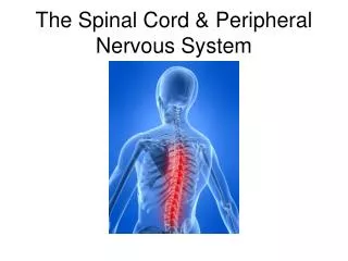
The Spinal Cord & Peripheral Nervous System. Peripheral Nerves. There are 12 pairs of cranial nerves and 31 pairs of spinal nerves. Spinal Nerves. There are 31 pairs of spinal nerves. All are mixed nerves (motor & sensory). Spinal Nerves. There are 31 pairs of spinal nerves.

E N D
Peripheral Nerves • There are 12 pairs of cranial nerves and 31 pairs of spinal nerves.
Spinal Nerves • There are 31 pairs of spinal nerves. • All are mixed nerves (motor & sensory).
Spinal Nerves • There are 31 pairs of spinal nerves. • All are mixed nerves. • There are: • 8 cervical spinal nerves (C8 comes out between C7 and T1) • 12 pairs of thoracic nerves, • 5 pairs of lumbar spinal nerves • and 5 pairs of sacral nerves.
Cervical plexus Cervical nerves C1 – C8 Brachial plexus Cervical enlargement Thoracic nerves T1 – T12 Intercostal nerves Lumbar enlargement Lumbar nerves L1 – L5 Lumbar plexus Sacral plexus Sacral nerves S1 – S5 Cauda equina Coccygeal nerve Co1
Cranial dura mater Terminus of medulla oblongata of brain Sectioned pedicles of cervical vertebrae Spinal nerve rootlets Dorsal median sulcus of spinal cord (b) Cervical spinal cord.
Anatomy of the spinal cord. Pia mater Epidural space (contains fat) Arachnoid mater Spinal meninges Subdural space Dura mater Subarachnoid space (contains CSF) Bone of vertebra Dorsal root ganglion Body of vertebra (a) Cross section of spinal cord and vertebra
Spinal Nerves The spinal nerve consists of a dorsal root and a ventral root. The ventral root contains efferent, motor neurons (voluntary and involuntary). The dorsal root is, afferent, sensory fibers.
Spinal Nerves The spinal nerve is only 1-2 cm long coming out from the spine and the branches into a dorsal ramus and a ventral ramus.
Dorsal root (sensory) Dorsal root ganglion Dorsal horn (interneurons) Somatic sensory neuron Visceral sensory neuron Visceral motor neuron Spinal nerve Ventral horn (motor neurons) Ventral root (motor) Somatic motor neuron Interneurons receiving input from somatic sensory neurons Interneurons receiving input from visceral sensory neurons Visceral motor (autonomic) neurons Somatic motor neurons
Ascending tracts Descending tracts Ventral white commissure Fasciculus gracilis Dorsal white column Fasciculus cuneatus Lateral reticulospinal tract Dorsal spinocerebellar tract Lateral corticospinal tract Rubrospinal tract Ventral spinocerebellar tract Medial reticulospinal tract Lateral spinothalamic tract Ventral corticospinal tract Ventral spinothalamic tract Vestibulospinal tract Tectospinal tract
Innervations of Specific Body Regions Except for the spinal nerves T2-T12, all other spinal nerves join into inter lacing networks called nerve plexuses. They are formed only by the ventral rami.
Dorsal ramus Ventral ramus Spinal nerve . Rami communicantes Intercostal nerve Dorsal root ganglion Sympathetic trunk ganglion Dorsal root Ventral root Branches of intercostal nerve • Lateral cutaneous • Anterior cutaneous Sternum (b) Cross section of thorax showing the main roots and branches of a spinal nerve.
Cervical Plexus This is formed from the first 4 cervical spinal nerves.
Cervical Plexus This is formed from the first 4 cervical spinal nerves. The most important nerve coming from this plexus is the phrenic nerve.
Cervical Plexus This is formed from the first 4 cervical spinal nerves. The most important nerve coming from this plexus is the phrenic nerve. This is the chief motor nerve for the diaphragm.
Ventral rami Segmental branches Hypoglossal nerve (XII) Ventral rami: Lesser occipital nerve C1 Greater auricular nerve C2 Transverse cervical nerve C3 Ansa cervicalis C4 Accessory nerve (XI) C5 Phrenic nerve Supraclavicular nerves
Cervical Plexus Damage to the spinal cord at C3- C5 could lead to respiratory arrest.
Brachial Plexus It is formed from C5-C8 and T1 with 5 major roots supplying the muscles of the muscles of the shoulder, thorax and upper limb.
Roots (ventral rami): C4 Dorsal scapular C5 Nerve to subclavius C6 Suprascapular Upper Posterior divisions C7 Trunks Middle C8 Lateral Lower Cords T1 Posterior Long thoracic Medial pectoral Medial Lateral pectoral Axillary Upper subscapular Musculo- cutaneous Lower subscapular Thoracodorsal Radial Medial cutaneous nerves of the arm and forearm Median Ulnar (a) Roots (rami C5 – T1), trunks, divisions, and cords Posterior divisions Roots Anterior divisions Trunks
Musculocutaneous nerve Lateral cord Axillary nerve Posterior cord Biceps brachii Coracobrachialis Medial cord Median nerve Radial nerve branches to triceps Radial nerve Ulnar nerve (b) Cadaver photo
Lumbrosacral Plexus It is formed from L1-L4 and lies within the psoas major muscle. The largest nerve of this plexus is the femoral nerve. The sacral plexus is formed from L4-S4 and lies just below the Lumbar Plexus
Ventral rami: Ventral rami Iliohypogastric L1 Ilioinguinal Femoral Lateral femoral cutaneous L2 Iliohypogastric Ilioinguinal Obturator L3 Genitofemoral Anterior femoral cutaneous Lateral femoral cutaneous Saphenous L4 Obturator Femoral L5 Lumbosacral trunk (a) Ventral rami and major branches of the lumbar plexus (b) Distribution of the major nerves from the lumbar plexus to the lower limb
Ventral rami: Ventral rami L4 Superior gluteal L5 Lumbosacral trunk S1 Inferior gluteal S2 Common fibular Tibial S3 Posterior femoral cutaneous S4 Pudendal S5 Sciatic Co1 Ventral rami and major branches of the sacral plexus
The largest nerve from this is the sciatic nerve. It is the largest and thickest nerve in the body. It supplies the entire lower limb. Irritation to this nerve gives rise to Sciatica.
Dermatomes `Dermatomes are areas of the skin innervated by the sensory nerves. These are fairly uniform and follow the nerve segments.
C2 C3 C2 C4 C3 C5 C6 C4 C7 C8 T1 C5 C5 T2 T1 T3 T2 T4 T3 T5 T4 T2 T2 T6 T5 T7 T6 T8 T9 T7 T10 T8 C6 C6 C5 C5 T11 T9 T12 C7 C7 T10 L1 C6 C6 S1 L2 C8 T11 C8 L3 S2 L5 L4 S3 T12 L1 L1 C6 S4 C6 S5 S2 C7 C7 C8 S3 C8 L2 L2 S1 S2 S2 S1 L1 L3 L3 L2 L5 L5 L4 L4 L3 L5 L5 L4 S1 S1 Anterior view (b) Posterior view L4 L4 L5 L5 S1
Reflex Activity • Reflexes can be inborn or learned. • An inborn reflex is a predictable, rapid response to a stimulus. • Acquired reflexes result from repetition.
Stretch and Golgi Tendon Reflexes The muscle spindles found in skeletal muscles provide information on the length of the muscle and the amount of tension on it. A common example of this is the patellar reflex. This reflex causes the muscle to contract in response to stretching.
The patellar (knee-jerk) reflex – a specific example of a stretch reflex 2 Quadriceps (extensors) 3a 3b 3b 1 Patella Muscle spindle Spinal cord (L2 – L4) 1 Tapping the patellar ligament excites muscle spindles in the quadriceps muscle. Hamstrings (flexors) Patellar ligament 2 Afferent impulses (blue) travel to the spinal cord, where synapses occur with motor neurons and interneurons. The motor neurons (red) send activating impulses to the quadriceps causing it to contract, extending the knee. 3a Excitatory synapse + Inhibitory synapse – 3b The interneurons (green) make inhibitory synapses with ventral horn neurons (purple) that prevent the antagonist muscles (hamstrings) from resisting the contraction of the quadriceps.
Golgi tendon reflexes Golgi tendon reflexes are the opposite and respond by causing muscle relaxation. This helps to prevent the muscle from over contracting.
1 2 Afferent fibers synapse with interneurons in the spinal cord. Quadriceps strongly contracts. Golgi tendon organs are activated. Interneurons Quadriceps (extensors) Spinal cord Golgi tendon organ Hamstrings (flexors) Efferent impulses to muscle with stretched tendon are damped. Muscle relaxes, reducing tension. Efferent impulses to antagonist muscle cause it to contract. 3b 3a + Excitatory synapse – Inhibitory synapse
Flexor and Crossed-Extensor Reflexes • These are initiated by painful stimuli and cause an automatic withdrawal of the threatened body part. Placing your hand on a hot stove is an example of this type of reflex.
+ Excitatory synapse – Inhibitory synapse Interneurons Efferent fibers Afferent fiber Efferent fibers Extensor inhibited Flexor inhibited Arm movements Flexor stimulated Extensor stimulated Site of reciprocal activation:At the same time, the extensor muscles on the opposite side are activated. Site of stimulus: a noxious stimulus causes a flexor reflex on the same side, withdrawing that limb.
Superficial Reflexes These are the result of gentle cutaneous stimulation. Common examples are the plantar reflex that tests the integrity of the corticospinal tract.
Babinski's sign This plantar reflex occurs when the corticospinal tract is damaged. In it the large toe dorsi flexes and the smaller toes fan laterally.
Spinal Cord Injuries Damage to the spinal cord results in paralysis (loss of motor function) or Paresthesia (sensory loss). These are devastating injuries since the cord will not heal.
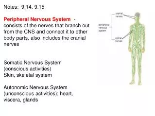
The Autonomic Nervous System The motor portion of the peripheral nervous system is divided into • the somatic (voluntary) motor system (SMS) and • the autonomic motor system (ANS).
Central nervous system (CNS) Peripheral nervous system (PNS) Figure 14.1 Place of the ANS in the structural organization of the nervous system. Sensory (afferent) division Motor (efferent) division Somatic nervoussystem Autonomic nervous system (ANS) Sympathetic division Parasympathetic division
Comparison The SMS stimulates skeletal muscle whereas the ANS innervates cardiac and smooth muscle as well as glands.
Comparison In the SMS the motor neuron cell bodies are located in the CNS and their axons extend to the muscles they innervate. It is a single neuron chain.
Comparison The ANS uses a two neuron chain. The first is the preganglionic neuron; its cell body resides in the CNS or spinal cord.
Comparison The ANS uses a two neuron chain. The first is the preganglionic neuron; its cell body resides in the CNS or spinal cord. The second motor neuron outside of the CNS. This second neuron is called the postganglionicneuron.
Neuro- transmitter at effector Cell bodies in central nervous system Effector organs Peripheral nervous system Effect Single neuron from CNS to effector organs ACh + SOMATIC NERVOUS SYSTEM Stimulatory Heavily myelinated axon Skeletal muscle Figure 14.2 Comparison of somatic and autonomic nervous systems. Two-neuron chain from CNS to effector organs NE ACh Unmyelinated postganglionic axon Ganglion SYMPATHETIC Lightly myelinated preganglionic axons + Epinephrine and norepinephrine ACh Stimulatory or inhibitory, depending on neuro- transmitter and receptors on effector organs AUTONOMIC NERVOUS SYSTEM Adrenal medulla Blood vessel ACh ACh Smooth muscle (e.g., in gut), glands, cardiac muscle PARASYMPATHETIC Lightly myelinated preganglionic axon Unmyelinated postganglionic axon Ganglion Acetylcholine (ACh) Norepinephrine (NE)
ANS Divisions The ANS is divided up into the parasympathetic and sympathetic divisions. They innervate the same organs and typically have opposite effects.