Download

1 / 18
180 likes | 201 Views
Explore intricate immune mechanisms & defence strategies with emphasis on lymphoid organs' histological structures. Covering nonspecific & specific defenses, immune cells involved, recognition of antigens, lymphocytes' role & more.

E N D
Immune-Lymphatic System - 1Introduction and Organisation 212 – 2005 – Week 8 Avinash Bharadwaj
Immunology : A vast and complex field • Defence mechanisms of the body • Perspectives • Molecular • Cellular • Histological • Gross anatomical • Emphasis : histological structure of lymphoid organs • Lecture 1 : • Simple concepts • Lymphatic nodules, Tonsil, Lymph node • Lecture 2 : • Spleen and Thymus • Further elaboration of immune processes
“Getting Rid Of ”… • External attackers • Viruses, bacteria, parasites… • Other “foreign” substances • Particulate matter and chemicals • Our own dead cells • Wayward cells • An unfortunate sidelight – our own normal cells
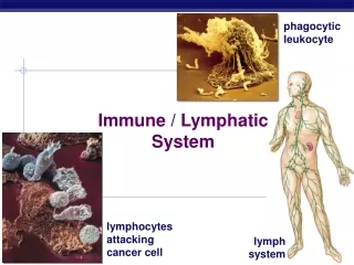
Mechanisms • Nonspecific defence • Specific defence • Cellular attack • Molecular (“humoral”) attack More than one mechanisms may be operating at a time! • Cells involved • White blood cells • Macrophages • Lymphocytes • Others • Intricate interactions between cells – molecular messengers
WBCs – Quick Review • Granulocytes • Cytoplasmic granules • Neutrophilic, eosinophilic or basophilic • Agranulocytes • Lymphocytes • Monocytes
Nonspecific Defence • Neutrophils • Movement out of capillaries • Phagocytosis and “digestion” • Expendable force • Action localised to site of infection • Other granulocytes…
Specific Defence - Immunity • Recognition of “non-self” or “foreign”. • Attack • Memory Terminology • Antigen – a foreign substanceOften (but not always) protein. • Antibody – a protein (γ-globulin) that specifically combines with an antigen.
Recognition of Antigens • The Antigen Presenting Cell • Macrophage • Phagocytosis • Fragmentation of foreign material • Attachment to cell surface molecules (MHC) • MHC (Major Histocompatibility Complex) • Proteins unique to each individual(Originally recognised in the context of tissue transplantation) • MHC Class I molecules present in all nucleated cells • MHC Class II – in APCs – Serve to bind antigens
Lymphocytes • Two major types – T and B lymphocytes • Not distinguished by LM • Two types of immune responses • Cell mediated – direct attack by cells • Humoral – antibodies as the medium (Humor : fluid. Antibodies are transported by the blood and also present in certain secretions))
Cellular and Humoral Immunity • “Cell mediated” immunity. • Cytotoxic T cells – Perforin, Lympotoxin, TNF • Helper T cells • Suppressor T cells • Memory • Humoral immunity • B lymphocytes plasma cells (abundant rER) • Memory B cells • Helper T cells
Exposure and Barriers • External surface – skin • The “open” systems • Digestive and Respiratory systems – most exposed • Urogenital (to a lesser extent) • The epithelial barrier • Integrity of epithelium • Intra-epithelial defence cells • Antibodies • “Local” infections and non-specific defence • Beyond the barrier…
Blood, Tissue Fluid and Lymph • Blood capillaries • “Extravasation” of fluid • Tissue fluid and exchange • Not all the fluid returns to blood vessels. • Lymph flows through lymphatic vessels before returning to larger veins.
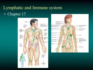
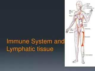
Lymphoid (Lymphatic) Tissues • Scattered lymphocytes and other cells • Lymphatic nodule – structural organisation • Lymphocytes, macrophages, plasma cells, other leucocytes • Outer zone (corona) and germinal centre • Aggregates of lymphatic nodules • Mucosa of GI Tract, respiratory system and other sites • Larger aggregates – Peyer’s patches (ileum), tonsillar tissue. • Organised, encapsulated structures • Lymph nodes • Spleen – special functions • Thymus – the academy
Lymphatic Nodule • “Non-capsulated” • Single or aggregated • Independent OR parts of other lymphoid organs • Outer dark zone • (Corona or cap) • Germinal centre
Tonsils • A group of lymphoid structures • Around the pharynx (GIT and RS) • Lingual, “palatine”, tubal, nasopharyngeal • Structurally similar • Epithelium of the pharynx • “Crypts” • Lymphatic nodules • Connective tissue and pharyngeal muscle.
Peyer’s Patches • Ileum – “antimesenteric border” • Lamina propria submucosa • Best seen in younger subjects Smaller aggregates present under many mucous membranes : “Mucosa Associated Lymphoid Tissue” or MALT
Lymph Node • Capsulated • Afferent lymphatics “subcapsular sinus” • Hilum – blood vessels, efferent lymphatic • Cortex and medulla • Cortex • Lymphatic nodules, germinal centres • “Paracortex” • Medulla • Medullary cords and sinusoids
C M More next week!