Competing Worldviews on Perception: Empiricism vs. Nativism in Brain Function Theories
530 likes | 651 Views
The study explores two competing theories of brain function regarding how perception is shaped: empiricism and nativism. By examining the roles of feedback and feed-forward processing in cortical microcircuits, the research highlights key findings from notable scientists, such as Sherrington and Hebb. It focuses on the control of postsynaptic calcium influx induced by neurotransmitters in the developing neocortex, emphasizing the crucial interactions between excitatory and inhibitory signals and their impact on synaptic activity and network dynamics.

Competing Worldviews on Perception: Empiricism vs. Nativism in Brain Function Theories
E N D
Presentation Transcript
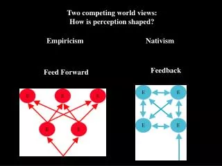
Two competing world views:How is perception shaped? Empiricism Nativism Feedback Feed Forward
Two competing theories of brain function: Feed forward: Sherrington Hubel &Wiesel Receptive fields Speed of processing Feedback: Brown Lorente/Hebb Llinás Recurrent connectivity Spontaneous activity
Neuron. 1991 Mar;6(3):333-44. Control of postsynaptic Ca2+ influx in developing neocortex by excitatory and inhibitory neurotransmitters. Yuste R, Katz LC. Laboratory of Neurobiology, Rockefeller University, New York, New York 10021. We assessed the pathways by which excitatory and inhibitory neurotransmitters elicit postsynaptic changes in [Ca2+]i in brain slices of developing rat and cat neocortex, using fura 2. Glutamate, NMDA, and quisqualate transiently elevated [Ca2%]i in all neurons. While the quisqualate response relied exclusively on voltage-gated Ca2+ channels, almost all of the NMDA-induced Ca2+ influx was via the NMDA ionophore itself, rather than through voltage-gated Ca2+ channels. Glutamate itself altered [Ca2+]i almost exclusively via the NMDA receptor. Furthermore, synaptically induced Ca2+ entry relied almost completely on NMDA receptor activation, even with low-frequency stimulation. The inhibitory neurotransmitter GABA also increased [Ca2+]i, probably via voltage-sensitive Ca2+ channels, whereas the neuromodulator acetylcholine caused Ca2+ release from intracellular stores via a muscarinic receptor. Low concentrations of these agonists produced nonperiodic [Ca2+]i oscillations, which were temporally correlated in neighbouring cells.Optical recording with Ca2(+)-sensitive indicators may thus permit the visualization of functional networks in developing cortical circuits.
Single-cell resolution imaging of Ca2+ influx due to action potentials • L5 pyramid loaded with 50µM fura • imaged by photodiode array at 1.6 kHz (0.6ms/frame)
Whole-cell filled AM filled
50 Hz 40 Hz
Cortical circuits in vitro are spontaneously active: spontaneous activity as a tool, let the circuit speak
Automatic identification of cells II/III IV V
700 500 Cell number 300 100 p < 0.05 * 7 6 5 % cells active / frame * 4 * * * * 3 * 2 1 0 Spontaneous synchronizations of a small % of neurons Low temporal resolution- 1sec/frame a
1 1 2 2 3 3 4 4 Synchronizations correspond to UP states -70 mV, 0 pA 9 mV 9 mV 9 mV 9 mV 1.3 s 500 ms 5 s 500 ms
Cortical motifs and songs: repeated sequences of activity Intermediate temporal resolution- 50 msec/frame
Repeated network activity measured in a single cell10 KHz resolution i iii iv
Repeated motifs of spontaneous activity in slices 10 pA 200 ms
Repetitions in vivo Ilan Lampl/David Ferster
What is role of thalamic stimulation on cortical dynamics? L2/3 L4 L5 Adapted from Brecht et al 2003
“Barrel” Cortex Thalamus Stimulation Electrode Imaging Layer 4 response to thalamic stimulation 4-8 stimuli 40 Hz 200 ms 50 – 100 mA Thalamic Stimulation
Thalamic stimulation generates cortical UP states • Prolonged depolarizations • ~ 10 mV depolarized from rest • Preferential state for action potential generation • Coincident with multiple nearby neurons 20 mV Vm -70 mV 1 s UP states
Spontaneous activity also generates cortical UP states 20 mV 500 ms Spontaneous
Spontaneous activity and thalamic stimulation engage the same neurons !!! Spontaneous Triggered Overlap X 5 X 4 Overlap Core Triggered Core Spontaneous Core
Similar Spontaneous and Evoked Intracellular UP states Overlap Spontaneous Triggered 5mV # of APs 20mV Amplitude 1 s 500 ms Duration Correlation of UPstates within cells Duration Amplitude No. APs
Identical Network Dynamics during Spontaneous and Evoked Network Events- 100 msec/frame Core Triggered Spontaneous 1 Frame Number Time 2 3
Millisecond Precision in the Repetition of Synaptic inputs during spontaneous and thalamic UP states 10 mV 500 ms 5 mV 100 ms
Interaction: Thalamic stimulation during spontaneous cortical UP states 10 mV
Mechanisms of spontaneous activity: pacemakers Spontaneously active neurons under synaptic blockade Control AP5/CNQX/PTX
Spontaneously active neurons under synaptic blockade 1 20 mV - 60 mV 20 s 2 AP5/CNQX/Picrotoxin 20 mV - 60 mV 20 s 3 20 mV - 63 mV 5 s
I I II/III II/III IV IV V V VI VI Type 1 Morphological characteristics of pacemaker cells “Thin” pyramidal morphology
2% DF/F 10 s Type 2 Second type of pacemaker cell
Type 2 Spontaneous oscillation in membrane potential. 1 -60 mV -67 mV 5 mV Frequency and amplitude of membrane potentials oscillations underlying burst firing were: 0.228 +/- 0.068 Hz 5.4475 +/- 1.89 mV 5 s 20 mV -67 mV 10 s 2 -61.5 mV -68 mV 5 mV 5 s 20 mV -68 mV 10 s
Type 2 Morphological characteristics I I II/III II/III IV IV V V VI VI Ascending interneuron morphology- Martinotti cell
Manipulating neuronal essembles 1) Stimulate multiple UP states: identify the early core 2) - Stimulate core to drive UP states - Or delete core to prevent UP states 2 Phot on cell Ablation APs Lots of Power Caged Glutamate
Controlling network activations: Does ablation of cells prevent UP states? Record pattern Ablate early-firing cells Blockade? ?
Pattern completion? ? ? Controlling network activations: Does the network complete patterns? Record pattern Stimulate to induce early observed sequence
Novel types of spontaneous and evoked cortical dynamics • Data: • • Reverberating activity is prevalent at all temporal scales • • Spatiotemporal patterns are real: statistics, two techniques, spatial profile, UP states, they can be triggered • Sparse dynamics: small number of cells • Single neurons can participate in many patterns • Repetitions never exact • Thalamic stimulation triggers internal states • Cortex is essentially “deaf” during spontaneous activity • Pacemaker cells in neocortex • Speculation: • Spatially organized ensembles: related to circuit features? • Preferred states: Hopfield attractors or metastable states? • Precisely repeated dynamics: Abeles’ synfire chains? • Sensory input reawakens internal cortical states • Cortex as a giant CPG?
Circuit attractors Input Attractors Inputs Memories Adapted from Wilson, 1999