Download
1 / 30
490 likes | 2k Views
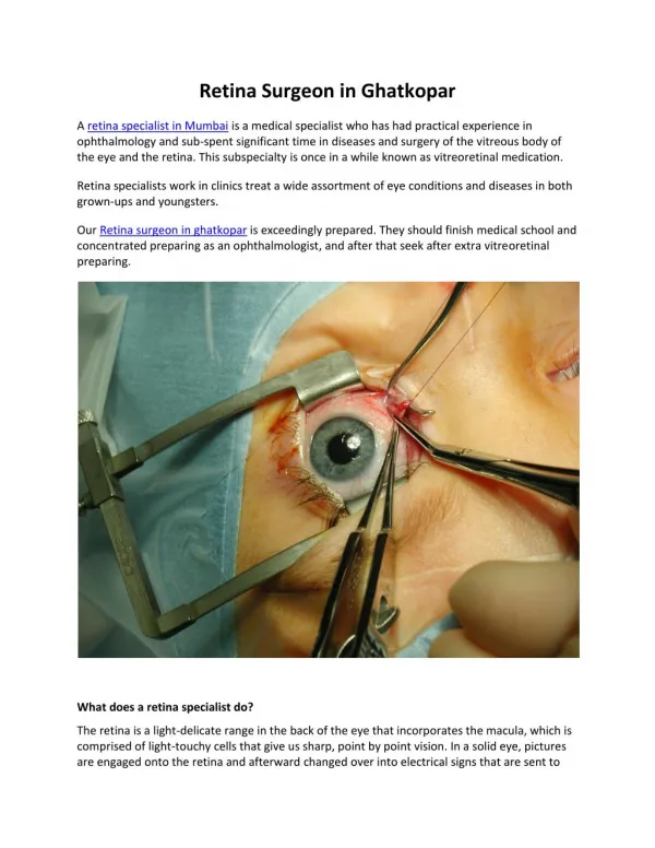
Vitreoretinal diseases (diseases of vitreous and retina). Anatomy of the EYE. anterior. posterior segment. Vitreous anatomy. Retinal anatomy. Retinal histology. Retinal histology. Macular anatomy. Retinal detachment- history.

E N D
Anatomy of the EYE anterior posterior segment
Retinal detachment- history • Gonin (1920) – primary role of retinal tear in retinal detachment
rhegmatogenous • tractional • serose Retinal detachment- classification
Rhegmatogenous RD Posterior vitreous detachment
Rhegmatogenous RD Retinal tear=(rhegma)
Rhegmatogenous RD Retinal tear=(rhegma)
Rhegmatogenous RD Pathological posterior vitreous detachment
Rhegmatogenous RD(upper quadrants) quickly progression
Rhegmatogenous RD(lower quadrants) slowly progression
Rhegmatogenous RD(symptoms) • Fotopsia (flashes) • Black spots (black snow) • Scotoma (black spot in peripheral visual field) • Decrease in visual accuity • Metamorfopsia
Rhegmatogenous RD(therapy) • Surgical • Extrabulbar operations (kryopexy,cerclage, plombage) • Intraocular operations (PPV with or without tamponade)
Kryopexy Development of chorioretinal adhesion (scar)
Pars plana vitrectomy (PPV) development of chorioretinal adhesion (endolaser) Retinal attachment (PFC)
Internal tamponade • Gas (SF6, C3F8) • Silicon oil Pars plana vitrectomy (PPV)
Pars plana vitrectomy (PPV) Gas tamponade
Pars plana vitrectomy (PPV) Silicon oil tamponade
Tractional forces Comparing of status before OP, after extrabulbar OP and after PPV
Tractional RD • can’t be find any tear • concave surface • can be detected epiretinal and subretinal membranes (diabetic retinopathy, trauma)
Tractional RD rhegmatogenous x tractional
Serose RD • can’t be find any tear • convex surface (high balloons) • free movement of subretinal fluid (tumors, uveitis, chorioidal detachment)
Serose RD Malignant melanoma of uvea Ultrasound of eye with MMU