Download
1 / 58
1.37k likes | 4.41k Views
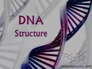
STRUCTURE OF DNA 1. primary structure 2. secondary structure 3. tertiary structure. PRIMARY STRUCTURE OF DNA. James Watson Francis Crick 1962 Nobel Prize. http://www.sciencemag.org/cgi/content/full/300/5617/255. http://www.lecb.ncifcrf.gov/~toms/icons/Watson.Crick.Nature.jpg.

E N D
STRUCTURE OF DNA1. primary structure2. secondary structure3. tertiary structure
James Watson Francis Crick1962 Nobel Prize http://www.sciencemag.org/cgi/content/full/300/5617/255 http://www.lecb.ncifcrf.gov/~toms/icons/Watson.Crick.Nature.jpg
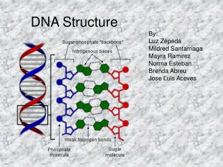
Polynucletide chain of DNA(primary/covalent structure) 1’ • The polynucleotide chain of DNA consist of nucleotides joined together by 3’,5’ phosphodiester bonds. • Phosphodiester bonds link the 3’- and 5’-sugar carbons of adiacent monomer. • Polynucleotides are directional macromolecules: each end of a polymer is distinct. • 3’ –end is one with a free 3’-hydroxyl. • 5’ –end is one with a free or phosphorilated 5’-hydroxyl. • Polynucleotides bear a negative charge at physiological pH 3’linkage 5’linkage
Simplified representation of DNA chain OH Sugar: horisontal line Base: single letter Phosphodiester bond: oblique line with P 3’ 5’ G P 3’ 5’ C P P 3’ 5’ T OH
Simplified representation of DNA chain Convention dictates that a single-stranded DNA sequence is written in the 5’ to 3’ direction. e.g. GCT means 5’GCT 3’
Quick Quiz Question : • Identify which is the 3’end of this oligonucleotide? • Write the simplified representation of this oligonucleotide.
Double helix of DNA • Watson and Crick discovered the secondary structure of DNA: a double helix– two sugar phosphate chains wrapping round each other, with the phosphate groups sticking out – the nucleotide from strand 1 meets the nucleotide from strand 2 in the middle. These pairs of nucleotides are complementary – where one strand has a C, the other has a G and vice versa; where one strand has an A the other has a T and vice versa. • Human DNA consists of approximately 3 x 10⁹ such “base pairs”.
The DNA double helix (secondary structure) DNA is double stranded: each molecule of DNA is composed of two polynucleotide chain that are joined together by formation of hydrogen bonds between the bases. DNA strands are twisted to form a double helix. DNA strands are antiparallel (one strand runs in the 5’ to 3’ direction and the other in the 3’ to 5’ direction, analogous to two street lanes carrying traffic in opposite direction).
The DNA double helix (secondary structure) 3. G-C pairs have 3 hydrogen bonds A-T pairs have 2 hydrogen bonds • One strand is the complement of the other, as they are formed by complementary bases (G is complementary to C, while A is complementary to T). • The concentration of deoxyadenosine (A) nucleotides equals that of thymidine (T) nucleotides (A=T), while the concentration of deoxyguanosine (G) nucleotides equals that of deoxycytidine (C) nucleotides (G=C)
Quick Quiz Question 1. Write the complementary strand of the following DNA strand: ATTTTAAGCTAAGGCCCTTT 2. Calculate the number of the hydrogen bonds existing beteen the complementary strands 3. Specify which base is at the 3’end of this strand and which base is at the 3’ end of its complementary strand.
The DNA double helix (secondary structure) • The B form of DNA described by Watson and Crick is right handed. • The distance spanned by one complete turn of the B-DNA double helix is 34 Å. • One turn of B-DNA includes 10 base pairs. • Oter forms of DNA include: - A-DNA which is more compact than B-DNA - Z-DNA is left handed and its bases are positioned more toward the periphery of the helix.
Quick Quiz Question • DNA • Is composed of nucleosides joined by phosphodiester bonds • Contains negatively charged phosphate groups • Contains base pairs A/T and G/C • Consists of two strands which run in the same direction.
What are the hydrogen bonds inside the DNA double helix? • Hydrogen bonds represent non-covalent (relatively weak) intermolecular bonds which are caused by the polarity of H-O bond.
From DNA to Chromosomes • In eukaryotes, the DNA is stored in the nucleus. Since there is not much space and DNA molecules are extremely large (the length of DNA from one singlehuman cell is 2m!!!), the DNA must be highly organised. • There are five levels of DNA compaction (or packing) that result in a 10.000 –fold decrease in DNA length.
Levels of packing of DNA in the nucleus-the first level- r • The DNA double helix is wrapped around protein complexes called histones- each unit of DNA wrapped round a histone complex is called a nucleosome. • Histones are small proteins, rich in basic aminoacids (Arg, Lys).
Levels of packing of DNA in the nucleus- the first level- r • Nucleosome contains an octameric core formed by two molecules of each histones H2A, H2B, H3, H4, around which 140 base pairs of DNA are wrapped. • The nucleosomes are separated by a small distance (DNA linker formed by 30 base paires). H1 histones interact with DNA linker. • The polynuceosome structure of DNA are similar to the “beads on a string” (=10nm fibrils of chromatin).
r “Beads on a string” appearance of DNA
r Levels of packing of DNA in the nucleus-the second level- • The polynucleosome chain is further compacted to form solenoid structures. • Solenoid structure has 6-7 nucleosomes pe turn. • Solenoid structure form the 30 nm fiber of chromatin.
r Levels of packing of DNA in the nucleus-the third, fourth and fifth levels- - The chromatin fibres fold together to form large looped domains. - These looped domains are then supercoiled and organised into distinct structures called chromosomes. - The human nuclear DNA ( genome) consists of 23 pairs of chromosomes.
The difference between diploid and haploid cells • Many eukaryotic cells contain pairs of chromosomes and are hence called diploid. • Other cells contain single chromosomes and are called haploid.
1 DNA
2 DNA DNA stands for deoxyribose nucleic acid This chemical substance is present in the nucleus of all cells in all living organisms DNA controls all the chemical changes which take place in cells The kind of cell which is formed, (muscle, blood, nerve etc) is controlled by DNA The kind of organism which is produced (buttercup, giraffe, herring, human etc) is controlled by DNA
3 DNA molecule DNA is a very large molecule made up of a long chain of sub-units The sub-units are called nucleotides Each nucleotide is made up of a sugar called deoxyribose a phosphate group -PO4 and an organic base
4 Ribose & deoxyribose Ribose is a sugar, like glucose, but with only five carbon atoms in its molecule Deoxyribose is almost the same but lacks one oxygen atom Both molecules may be represented by the symbol
5 The bases Adenine (A) Thymine (T) Cytosine (C) (G) Guanine The most common organic bases are
6 Nucleotides PO4 adenine deoxyribose The deoxyribose, the phosphate and one of the bases Combine to form a nucleotide
Joined nucleotides PO4 PO4 PO4 PO4 sugar-phosphate backbone + bases 7 A molecule of DNA is formed by millions of nucleotides joined together in a long chain
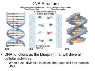
8 In fact, the DNA usually consists of a double strand of nucleotides The sugar-phosphate chains are on the outside and the strands are held together by chemical bonds between the bases
2-stranded DNA PO4 PO4 PO4 PO4 PO4 PO4 PO4 PO4 PO4 PO4 PO4 PO4 PO4 PO4 PO4 PO4 9
10 Bonding 1 Adenine Thymine Guanine Cytosine The bases always pair up in the same way Adenine forms a bond with Thymine and Cytosine bonds with Guanine
11 Bonding 2 PO4 PO4 thymine adenine PO4 PO4 cytosine guanine PO4 PO4 PO4 PO4
12 Pairing up PO4 PO4 PO4 PO4 PO4 PO4 PO4 PO4 PO4 PO4 PO4 PO4 PO4 PO4 PO4 PO4
13 The paired strands are coiled into a spiral called A DOUBLE HELIX
14 THE DOUBLE HELIX bases sugar-phosphate chain
15 A DIY model ofpart of a DNA molecule
16 replication During a cell divides, the DNA strands unwind and separate Each strand makes a new partner by adding the appropriate nucleotides The result is that there are now two double-stranded DNA molecules in the nucleus So that when the cell divides, each nucleus contains identical DNA This process is called replication
17 The strands separate PO4 PO4 PO4 PO4 PO4 PO4 PO4 PO4 PO4 PO4 PO4 PO4 PO4 PO4 PO4 PO4
PO4 PO4 PO4 PO4 PO4 PO4 PO4 PO4 PO4 PO4 PO4 PO4 PO4 PO4 PO4 PO4 PO4 PO4 PO4 PO4 PO4 PO4 PO4 PO4 PO4 PO4 PO4 PO4 PO4 PO4 PO4 PO4 18 Each strand builds up its partner by adding the appropriate nucleotides
Genetic code 1 19 The sequence of bases in DNA forms the Genetic Code A group of three bases (a triplet) controls the production of a particular amino acid in the cytoplasm of the cell The different amino acids and the order in which they are joined up determines the sort of protein being produced
Genetic code 2 Ser-Cyst-Val-Gly-Ser-Cyst Ala Val Val-Cyst-Ser-Ala-Ser-Cyst-Gly Val- Cyst-Ala-Ala-Ser-Gly 20 This is a small, imaginary protein molecule showing how a sequence of 5 different amino acids could determine the shape and identity of the molecule Each amino acid (Serine, Cysteine, Valine, Glycine and Alanine) is coded for by a particular triplet of bases
Coding Cytosine Adenine Thymine 21 For example Valine Codes for Cytosine (C) Alanine Codes for Guanine (G) Adenine (A)
Triplet code 22 This is known as the triplet code Each triplet codes for a specific amino acid CGA - CAA - CCA - CCA - GCT - GGG - GAG - CCA - Ala Val Gly Gly Arg Pro Leu Gly The amino acids are joined together in the correct sequence to make part of a protein Ala Val Gly Gly Arg Pro Leu Gly
23 DNA and enzymes The proteins build the cell structures They also make enzymes The DNA controls which enzymes are made and the enzymes determine what reactions take place The structures and reactions in the cell determine what sort of a cell it is and what its function is So DNA exerts its control through the enzymes
Genes 24 A sequence of triplets in the DNA molecule may code for a complete protein Such a sequence forms a gene There may be a thousand or more bases in one gene
Question 1 Which of the following are components of nucleotides? (a) deoxyribose (b) amino acids (c) phosphate (d) enzymes (e) organic bases